


The literature analysis suggestes that a complex characterization of physiological activity of a living organism requires dozens or hundreds of chips depending on the task. For example, there are both active and passive chips used in biophysical, molecular metabolomic and genomic studies in fundamental and applied molecular medicine:
a. allergology (Taira, 2009; Lupinek, 2014; Seyfarth, 2014; Zienkiewicz, 2014; Williams, 2016);
b. haematology and transfusiology (Hassan, 2015; Nguyen, 2015; Chen, 2015; Kuan, 2015; Rafeie, 2016; Mielczarek, 2016), including blood-brain barrier research / modeling (Shao, 2016; Bonakdar, 2016; Brown, 2015; Deosarkar, 2015);
c. lymphology (Hanna, 2003; Shimizu, 2007; Moura, 2016) and phlebology (Franco, 2012; Brivo, 2012; Zhou, 2012; Ryu, 2015);
d. cardiology (Tanaka, 2007; Chean, 2010; Grosberg, 2011; Agarwal, 203; Wang, 2014; Rismani, 2015; Jastrzebska, 2016; Marsano, 2016; Zhang, 2016);
e. gastroenterology-on-a-chip (Yang, 2009; Esh, 2012, 2014), including gut-on-a-chip techniques (Bjerketorp, 2008; Kim, 2008, 2016; Tottey, 2013; Lee, 2016);
f. cellular neurophysiology-on-a-chip and neuromorphogenesis-on-a-chip (Millet, 2010; Ling, 2010; Kim, 2014; Huang, 2014; Wei, 2014; Kunze, 2015; Yamada, 2016);
g. endocrinology (Marchesini, 2007; Bovet, 2007; Srivastava, 2014);
h. immunology (Yakovleva, 2002; Yang, 2005; Corgier, 2007; Liu, 2011; Zhang, 2011; Kayo, 2013; Wang, 2015; Ali, 2016);
i. general “splanchnology-on-a-chip” based on N principally equivalent approaches: “organ-on-a-chip” (Wikswo, 2013; Ahmad, 2014; van der Helm, 2016; Mousavi, 2016) / “organ-on-a-chip” (Lee, 2013; Bhise, 2014; Odjik, 2015; Kim, 2015; Caplin, 2015; Sticker, 2015; An, 2015; Zheng, 2016; Cho, 2016), “organoid-on-a-chip” (Skardal, 2016) and “physiome-on-a-chip” (Stokes, 2015), which can be integrated in the frame of concept “body-on-a-chip” (Esh, 2011, 2016; Williamson, 2013; Reif, 2014; Sung, 2014; Kelm, 2014; Ryu, 2015; Perestrelo, 2015); {etc.}
The above problem made the study so complicated, that it became quite unfeasible, since the “multi-chip” analysis (see Terminological remark No. 1) turned to be very expensive and the large sample volume required for such a complex analysis could not satisfy the principles of non-destructive diagnostics on a chip (for example, see (Takahashi, 2004; Feng, 2015)) due to many biomaterial sampling points (for example, see (Ando, 1987; Nikolaidis, 2012)) standardized in the protocols for biomedical and veterinary diagnostics.
On the other hand, the difference and variety of the sampling and the sample preparation techniques for different microchips and standard diagnostic methods made the problem of analyzing the complex biochemical physiological state of the organism unimplementable and poorly informative. It is quite obvious that for the purpose of compatibility and comparability of the measurement using different analytical devices (see Terminological note No. 2) it is necessary to provide the compatibility and comparability of the sampling and the sample preparation methods. In the ideal case, all the analytical procedures should be performed with a single uniformly calibrated device using the same sample for all the tests without moving the sample from one device to another. To date there are independent calibration methods for chips (Gillot, 2007; Binder, 2008; Karsunke, 2009; Nakamoto, 2010; März, 2010; Song, 2012; Buchegger, 2014), as well as the calibration protocols for other analytical methods (including the imaging ones) using chips (Su, 2016; Garnica-Garza, 2009). Hence, we need an equivalent of cross-calibration in the interpretation close to that given by NIST for cytometry (Hoffman, 2012), although the term was used much earlier in radiology (including tomography) and nuclear medicine (Paans, 1989; Genant, 1994; Tothil, 1995; Grampp, 2000; Geworski, 2002; Hetland, 2009; Garnica-Garza, 2009), as well as in the number of spectroscopic methods applied for the biomaterial analysis (Kwiatkowska, 2008; Wang, 2012; Poto, 2015; Liu, 2016).
In addition, when we deal with the structured samples such as biological tissues, it is also important to obtain information on the spatial distribution of the substance or property analyzed in the image form, for example:
o magnetic field imaging;
o electrochemical parameters and field gradient;
o laser beam transmission outside the visible spectral range;
o distribution of the emitting regions in autoradiography;
o polarization characteristics and the angular fluorescence polarization;
o the local temperature of the sample at different points on a chip{etc.}
Moving the sample from one microscope to another makes it difficult to establish the correspondence (colocalization) between the regions of interest (ROI) for different wavelength ranges (or different physical characteristics) allowing to perform the mapping and identification of the components under investigation due to the difference of visualization in different spectral ranges (or different physical “descriptors”). This prevents one from combination of the signal distribution maps from different spectral regions, and hence, makes it impossible to establish the correlations between the presence and distribution of the certain components or physical and chemical properties in the sample / tissue.
Since different components of the analyte possess a number of colocalized characteristics in different spectral ranges (Zimmermann, 2005; Gavrilovic, 2009), it is possible to perform either a simultaneous or a sequential mapping and identification of several tissue components based on the physically different properties. For example, some target components can be visualized using non-spectral properties, such as magnetic fields (Gruschke, 2012; Kim, 2015; Hejazian, 2015), labeled atom diffusion (for example, see: Parker, 1981; Galbraith, 1981; Blakely, 1986; Hein, 1986; Nemecz, 1988; Pouteau, 2003), temperature maps (Choudhury, 2012; Rosenthal, 2014; Karadimitriou, 2014; Meng, 2015; Lo, 2016) or redox maps (including ratiometric those (Herman, 2005; Hilderbrand, 2008; Zhang, 2015; Chen, 2015; Pan, 2016)) on a chip (Jezierski, 2013; Gashti, 2016). We propose to implement a full range of methods for mapping the biological tissue parameters with or without specific labels using planar transducers / converters of the non-optical signal to the optical one, as will be described below.
This will also result in the substitution of a number of independent expensive diagnostic devices with a simple unified complex diagnostic and analytical device. The operator of such a complex lab-on-a-chip will predominantly perform data analysis and processing (a so-called data mining, which is now mainly used not in the active mapping or imaging chips, but in the passive chips for genomic and peptidomic investigations (Lee, 2001; Smith, 2005; Abascal, 2008; Ghanekar, 2008; Usui, 2009; Nussbeck, 2013)) rather than routine analytical procedures (such as sampling and dropping (Fang, 2002; Du, 2005; Cellar, 2005; Huynh, 2006; Zhang, 2007; Do, 2008; Jang, 2009; Kertesz, 2010; Sun, 2010; Coskun, 2010; Wu, 2012)) due to an automatic machinery. This is in consistence with the modern trends in the development of the information society and the extension of the applicability of the chemoinformatic (“chemobioinformatic” (Basak, 2012)) software for biomedical and pharmaceutical (Weinstein, 2001; Shedden, 2003; Shedden, 2004; Parker, 2004; Ghose, 2006; Kong, 2008; Speck-Planche, 2014; Capasso, 2015; Gromova, 2016), agrobiological and biotechnological problems (Speck-Planche, 2012; Grädow, 2014).
In this regard, the design of the above proposed complex devices for multi-parametric analysis and mapping of the samples is of great importance for analytical practice both for improving the quality and information content of the analysis and for the rational use of the working time of the analyst. The possibility of connecting such devices to the PC and mobile network resources (Lillehoi, 2013; Wu, 2014; Pan, 2014; Koydemir, 2015; Bhavnani, 2016) allows to improve the quality of telemedicine (Fleck, 1999; Bishara, 2011; Balsam, 2015), GIS – coupled analysis / sample analysis in the field conditions with the geodetic reference (Senbanjo, 2012 Gerald, 2014; Ferguson, 2016), quality control on a chip (Shearstone, 2002; Hartman, 2005; Zhang, 2005; Stokes, 2007; Pierzchalski, 2012) in chemical and biotechnological industry using SCADA and similar systems (Gieling, 1996; Ozdemir, 2006; Smith, 2006; Moya, 2009).
The implementation of the technology proposed will increase the labor productivity of the analysts and researchers, since the performance of N analyses with a single device equals to the N-times reduction of the amount of the auxiliary routine work compared to the performance of each analysis with an independent device requiring different sampling procedures and sample treatment protocols. Since the first labs-on-a-chip were developed by the author for his own research problems and were tested in the routine research practice, he could easily appreciate the ergonomics and usability of such devices with the maintenance of the quality and increase in the rapidity of the analysis.
Novel approach
The contemporary analysis of the literary and the preliminary calculations, suggested using an optical channel for analytical data acquisition with the CMOS and CCD detectors. However, the serial CMOS and CCD allow detection only the optical parameters providing the analyte concentration measurements by absorbance or transmittance or fluorescence of a selectively bound dye. Modern CMOS- and CCD-based labs-on-a-chip fail to perform visualization of a number of characteristic descriptors for many biological and medical samples, such as magnetic fields, temperature profiles, localization of radioisotope sources and selective emission from cells and tissues in autoradiography, etc. Meanwhile, nothing prevents us from using the primary signal converters of the required parameters / variables into the optical signal.
There are known:
· magnetooptical converters and indicator films (Anderson, 1968; Harms, 1980; Aulich, 1980; Papp, 1980; Arkhangel’skii , 1986, 1989; Challener, 1987; Mao, 1989; Challener, 1990; Krafft, 2004; Fratello, 2004);
· radiation-optical (spectro-)colorimetric converters (Apanasenko, 1981; Kulagin, 1983, 1984, 1985, 1987; Bazylev, 1992; Mikhailov, 1996; Kulagin, 2003, 2006; Kulagin, 2007; Sadulenko, 2009) and thin film scintillators (Albul, 1968; Avdeyev, 2001; Garcia-Murillo, 2003; Berdnikov, 2013; Tolstikhin et, 2014; Inami, 2015; Rincón-López, 2016; Park, 2016);
· thermo-optical effect transducers-converters (Malashko, 1974; Dolgov, 1979; Pálfalvi, 2004; Liberts, 2005; Gunyakov, 2006; Nedosekin, 2007; Loiko, 2012), including thermochromic ones (Soloway, 1955; Chivian, 1972; Yang, 1979; Mazumder, 1995; Qazi, 2003; Siegel, 2009; Sia, 2009; Shelton, 2010; Qian, 2012; Heo, 2012; Zhou, 2013; Funasako, 2013; Li, 2013; Bond, 2013; Seeboth, 2014; Kim, 2014; Wan, 2015; Liu, 2016; Zhang, 2016), including infrared-sensitive metamaterials;
· chemo-optical active interfaces (van Gent, 1990; Wroblewski, 1997), colorimetric or flouorimetric indicator films (Chen, 1997; Nakamura, 2003; Kowada, 2004; Lü, 2006; Thomas, 2009; Gao, 2011; Kassal, 2014; Mills, 2016; Choi, 2017) and papers (Yeoh, 1996; Ostrovsakaya, 2004; Gaiduk, 2009; Ganesh, 2014);
· electroluminescent (Vlasenko, 1966; Shaposhnikov, 1970; Ramazonov, 1972; Samokhvalov, 1993; Brigadnov, 1993; Gurin, 1997; Savikhib, 1997; Zabudskii, 2000; Maltsev, 2011; Rodionov, 2013; Meshkov, 2014; Evsevichev, 2016) and cathode-luminescent indicators / phosphors (Tebrock, 1968; De Mets, 1971; Suzuki, 2009; Obraztsov, 2013; Kaz, 2013; Shi, 2014; Li, 2016)
and other position-sensitive target signal converters into the optical signal[1], which allow a direct realization of the “two-level conversion” including a first conversion of the analytical signal into the optical one by the planar converter located above the photosensitive CMOS / CCD detector with the subsequent conversion of the optical signal into the electrical one by the optoelectronic converter (CMOS or CCD). The above converters being placed into the cartridge or cassette system, or the rotating disc (this is a reversible idea from lab-on-a-disc design (Park, 2012; Glass, 2012; Hwang, 2013; Bosco, 2013; Delgado, 2016)) can be replaced by one another in real time allowing to vary the measuring parameters, and hence, providing the sequential mapping and measuring of the above parameters.
At the first step the single devices (chips and the corresponding readers) have been developed for the single parameter registration (e.g. a special compact device for magnetic field visualization has been designed using the magnetic film converter (flux detector) and a similar radiographic visualizer has been developed based on the scintillation plates). Later these devices were combined into a single hybrid device with the incomplete set of the primary converters for the purposes of the complex analysis (see Figures 1-3). At the final step we are going to overcome those limitations and to develop a hybrid multi-functional lab-on-a-chip allowing to perform in a single run of the cassette with the cartridges-converters the full position-sensitive mapping of the spatial distribution of the following parameters:
I. spectral / colorimetric, densitometric and fluorescent parameters of the analyte for histochemistry and immunofluorescent analysis;
II. luminosity distribution beyond the optical spectral range for laser diagnostics or the on-chip LDV, LDA, LDF, laser-accisted PIV;
III. magnetic field for selective staining of biological tissues with the magnetic nanoparticles or for the on-chip testing of the pharmaceuticals' targeting in the external field;
IV. distribution of the emitting regions in autoradiography and for the sample analysis with the radioactive contamination;
V. polarization parameters and the fluorescence polarization for those cases when the rotation of the polarization plane is a diagnostic criterion, from simple saccharimetry to the chirality-based analytical methods introduced from molecular biology;
VI. the slide temperature (for the living slices and tissue cultures) for determination of the biothermogenesis intensity or the redox transformation intensity, which is one of the most important diagnostic criteria of the neoplastic processes in biopsy;
VII. pH, Eh, pX, etc. using discrete indicator films by the colorimetric, spectrocolorimetric or fluorescence response signal (see Figure 4).
The cartridges-converters can be either built into the chip reader (the most suitable configuration for the ultracompact disposable chips without the recording and processing units) or implemented directly into the autonomic chips in the case of the autonomous reusable devices. In the early prototypes developed by the author the chip was combined with the reader forming a so-called self-reading chip capable of the telemetric data translation through a radiofrequency channel (Notchenko, 2012, 2013).

Figure 1. The first version of the multiparametric reader system for multidescriptor mappimg of biological samples.

Figure 2. The second version of the multiparametric reader system for multidescriptor mappimg of biological samples.
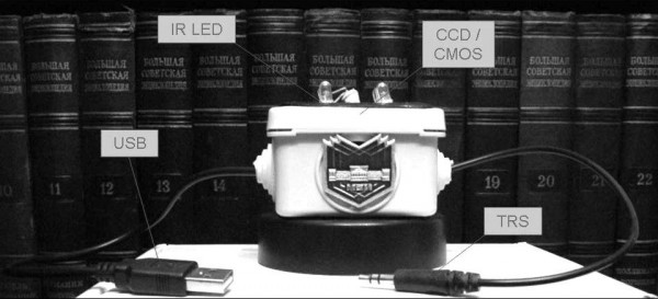
Figure 3. The ultracompact lab-on-a-chip reader with USB and TRS.

Figure 4. Redox-signal conversion into the optical signal using colorimetric / spectrocolorimetric “chemical pixels” (“chemical resells” / “sensels”): A,C – baseline (reactive film without detective dye response); B,D – indicator signal. Equivalent mosaic sensor-converter elements may be assembled using different converters and indicators (not only “pH-pixels” / “pХ-pixels”, but also “electric luminescence pixels”, “magnetic field pixels”, “radiation pixels”).
Preliminary results
To date there are three types of the prototype of the device proposed above:
A. a compact chip fabricated using the radioelectronic methods, which requires a special compact reader also developed by the author (Figure 1; Figure 2);
B. a compact lab-on-a-chip with the built-in system of converters and a broadcasting system (Figure 3);
C. a laser lab-on-a-chip with the rotating cassette and a disc.
There is also a submersible (soil / marsh (Gradov, 2012)) sealed topology of the device, fabricated using a 3D printer and capable of functioning in the soil environment for several months and broadcasting the signal to the remote wireless telemetry receiver (Notchenko, 2013).
Further development of this principle includes the selection of the optimal converters for different forms of visualization, the search for the most effective and selective filters, high-resolution and the most sensitive detectors, the device specialization and testing for different analytical tasks and sample types. It is potentially feasible to design a number of film converters (ferroelectric and piezoelectric (Hussain, 1971; Moyle, 1989; Shih, 1998; Bu, 2007; Wang, 2009; Cao, 2012; Sanada, 2015; Alluri, 2015), pyroelectric (Brown, 1989; Lehman, 2000, 2007) etc.) which can be implemented into the complex device after the optimization technology. These aims will result in the design of a highly complex multifunctional analytic device for a wide range of applications.
With the reduced number of converters this device can be specialized to any local task and research area being modified and completed according to the certain customer requirements (customization). For example, in biomedical laboratories it is expected to be provided only with the fluorescent, thermographic and polarization-sensitive cartridges (analogically to widely-used different complex AutoAnalyzers (Moyles, 1982; Jeie, 1983; Shipley, 1990; Billet, 1994; Fleming, 2001)), while for nuclear chemistry on a chip (De Leonardis, 2011; Arimaet, 2014; Rensch, 2014) and radiation material studies the scintillation converters with different quenching factors are expected to be the most suitable.
The Open Source software (with the SCADA-like GUI interface (Mercurio, 2009)) for our Open Hardware (Powell, 2012; Fisher, 2015) should also be developed for such multiparametric devices in order to enable an easy adaptation of the measurement scheme in accordance with the needs of each user by means of writing an individual module for the control of switching between the primary cartridges-converters with the corresponding shift of scales and measurement / visualization units.
It is also possible to perform the correlation distribution analysis of any other sample characteristics derived from the above listed parameters (e.g. redox potential from the fluorescence measurements using specific dyes or the heat capacity from the thermographic data), i.e. colocalization analysis (Adler, 2007, 2008, 2010; Chen, 2011). Thus, we propose a novel analytical method using labs-on-a-chip capable of the complex analysis of various analyte parameters, provided by the design of a cassette with different converters moving relative to the sample.
Examples of applications of the prototypes
A. Monitoring of the biophysical parameters of cells and tissues during morphogenesis of the complex structures in a spectrozonal / multispectral mode, as well as at various angles with the polarization analysis (Notchenko, 2013) controlled by the five-axis automatic system.
B. Synchronous in situ studies of the fluorescent characteristics of the neuron development and their electrophysiological activity (Zaytsev, 2014; Alexandrov, 2015).
C. Cyclic code decoding of the nucleic acids including xenonucleic acids (Orehov, 2014) and DNA-cryptography; genetic data qualimetry for synthetic biology, as well as paleogenetic and molecular phylogenetic data using a chip-sequencing technique (Gradov, 2014).
D. The studies of colocalization of not only the biochemical agents, but also of their systems biological (SBGN), physico-chemical (QSPR), biophysical and pharmacological (QSAR) descriptors, derived from the automatic computer interpretation of the analytical data (COBAC) from the chip (Orehov, 2015, 2016).
E. Redox-mapping, including the study of the reactive oxygen species localization using lab-on-a-chip for ozonometric microscopy (Gradov, 2013). Any positional-sensitive measurements in a lab-on-a-chip can be calibrated by the spectrum using spectrophotometric / colorimetric temperature for the tuple chemometric analyte systematization (Gradov, 2014), as well as by the spatial coordinates for morphometric purposes and colocalization studies using different counting chambers’ grids (Gradov, 2012) with the chamber microgrooves serving as a useful analytical microfluidic instrument.
F. The studies on the reaction-diffusion processes coupled to the redox reactions which simulate primitive morphogenesis in biomimetic heterogeneous media, accompanied by the oscillatory and autowave behavior of the active medium localized on the chip (Gradoff, 2012), particularly under optical pumping (Gradov, 2015) with the appropriate filter in a mechanically controlled cartridge cassette of the chip.
G. Real-time monitoring in microbiological studies of the soil, greenhouse and wetland environments (Gradov, 2012) with the telemetric radiofrequency signal transduction, which substitute the classical soil chambers and Rossi-Cholodny slides, providing a direct in vivo and in situ monitoring of the microbial community parameters in a telemetric mode instead of the subsequent analysis after the removal from the natural environment. The above system with the RF broadcasting for the soil biophysical and microbiochemical monitoring is similar to the telemedicine of the future for agricultural industry.
H. The possibilities of the above analytical technique have been significantly expanded in recent years due to the development of the novel data processing software which allows to study the size distribution of the quantum dots, to perform the nucleic acid code decoding and to use the lab-on-a-chip as a spectrometric system with a complicated signal processing using a number of different methods in addition to the simple Fourier transform.
Thus, it is possible to develop a so-called multifactor “lab-on-a-chip on demand”, which in its basic version will include: fluorescent analyzer; mapping polarimeter; thermograph (thermovisiograph); IR- and UV-range visualizer; magnetograph; scintillation visualizer “spinthariscope-on-a-chip” for autoradiography; “cymatic analyzer” based on a thin piezoelectric element and CCD / CMOS registrator.
Supplement
Terminological remark No 1
Although, strictly speaking, “multi-chip” / “multichip” application in microfluidics is not fully correct and conventional, since their area of biological application is limited to the retinal stimulation devices (Tokuda, 2009, 2010) and photobionic / photobiomimetic retinal models like “Analog Silicon Retina” (Kameda, 2006), including those with the visual orientation emulation (Shimonomura, 2005), as well as in neurophysiological modeling and recording (Vogelstein, 2007; Gosselin, 2009). In other cases, multichips are predominantly used in light engineering (Christensen, 2002; Chien, 2007; Kim, 2010; Oh, 2011; Horng, 2012; Shubin, 2015) and optoelectronics (Gruber, 2004; Milojkovic, 2006; Fan, 2006; Nazarathy, 2006). Moreover, this term originated in the early 1990-th, long before the emergence of the microfluidic trend (Zaleta, 1994; Fan, 1995; Zhao, 1997; Cruz-Rivera, 1998; Haney, 1999). The appearance of the term “multichip” in the works on microfluidics is sporadic and random compared to the above cited papers. This term once appears in the title of the work on PCR-chip (Panaro, 2004) and another time – in the paper concerning the on-chip analysis of the pharmacological formulations (Al Lawati, 2011). It is even more correct to speak about the “multi-sensor chip” / “multisensor chip” (Abramova, 2009; Sellami, 2010), which is a consequence of the development of the microminiaturization trend (“multisensor arrays”) since 1990-th (Mandenius, 1998; Bachinger, 1998; Paulsson, 1999) to the present time (Sharpe, 2014). However, it should be noted that a multi-sensor chip by definition possesses not a single sensor with multiple tranducers providing one-to-one correspondence between each of the converted physical quantities and the sensor response depending on the transducer type, but a number of sensors based on different physical principles and calibration parameters, with the separate task of the obtained data acquisition and processing. Therefore, in chemometrics multitranducer chips / multitransducer sensors and multitransducer arrays are mostly applied not for the position-sensitive measurements (Kurzawski, 2006; Jin, 2008). In the ultrasonic studies which require scanning, the meaning of the term “transducer” differs from that in analytical chemistry (Pride, 1974; Pedersen, 1977; Fessenden, 1984), but the scanning process in most cases is performed with the assistance of the operator, except the photoacoustic tomography and other tomographic methods (Deng, 2016; Lindsey, 2011), while in them the position sensitivity is usually provided by the external scanning system with the stepper motors rather than by the sensor or transducer properties.
Terminological remark No 2
“Cross-method convergence”, “cross-method assessment” or “cross-method compatibility” – a standard requirement for multi-method biomedical studies (Pavot, 1991; Meyer, 1996; Turner, 2006; Chou, 2011; Handelzalts, 2014). Regarding labs-on-a-chip, analytical microchips and microarrays, cross-method studies are rare. In most studies the data compatibility from the chips is considered only as the “cross-platform” or “cross-laboratory” requirement (especially in genomic, transcriptomic and translatomic studies (Stafford, 2007; Sumida, 2007; Liu, 2008, 2013; Vermeulen, 2009; Mistry, 2010; Jiang, 2010; Gonen, 2015; Foster, 2015)), instead of the comparison by a number of variables between the data obtained from different sensors (e.g. concentration) at a single chip. Consideration of the data compatibility from different chips or different sensors of a single chip as the descriptor correlation is not performed automatically in microfluidic analytic devices, although correlation spectroscopy of the process patterns on a chip (Bougot-Robin, 2012; Travagliati, 2013), as well as on-chip correlation fluorescence spectroscopy (Rudenko, 2009; Chen, 2011) are widely applied without consideration as the system state descriptors on a chip. Such innovation could be also applied for cross-calibration in the interpretation close to that given by NIST for cytometry (Hoffman, 2012), although the term was used much earlier in radiology (including tomography) and nuclear medicine (Paans, 1989; Genant, 1994; Tothil, 1995; Grampp, 2000; Geworski, 2002; Hetland, 2009; Garnica-Garza, 2009), as well as in the number of spectroscopic methods applied for the biomaterial analysis (Kwiatkowska, 2008; Wang, 2012; Poto, 2015; Liu, 2016).
Acknowledgements
This work is supported by RFBR (Project No. 16-32-00914).
[1] See, for example, a review “Unusual effect colourants” (Gregory, 2003): “An unusual effect colourant is one that exhibits a colour change or some other unusual effect outside the traditional colour-imparting properties of a colourant. Il also includes novel ways of producing colour. Many such effects are known and commercialised. For example, holograms and optically-variable pigments, which utilise the interference of visible light, and the electrostatic and photoconductive effects used in photocopiers and laser printers. However, this paper focuses on effects involving dyes and pigments either directly or indirectly for the unusual colour effect. These effects may be conveniently classified under two headings: Luminescent effects; Chromisms (colour change effects). Luminescent effects include fluorescence, phosphorescence, twisted intramolecular charge transter (TICT) states, electroluminescence, both of small molecules and polymers, chemiluminescence, bioluminescence and iridescence. The chromisms include the relatively familiar ones such as photochromism, thermochromism and electrochromism, plus less familiar ones including barochromism (colour change with pressure), chronochromism (colour change with time), and even claustrophobic dyes! The unusual effect may be caused by a single colourant or a composite system. Both types will be exemplitied”. (JAG).
References
1. Abascal F, Carmona-Saez P, Carazo JM, Pascual-Montano A. ChIPCodis: mining complex regulatory systems in yeast by concurrent enrichment analysis of chip-on-chip data. Bioinformatics 2008, 24(9):1208-1209.
2. Abramova N, Ipatov A, Levichev S, Bratov A. Integrated multi-sensor chip with photocured polymer membranes containing copolymerized plasticizer for direct pH, potassium, sodium and chloride ions determination in blood serum. Talanta 2009, 79(4):984-989.
3. Adler J, Pagakis SN, Parmryd I. Replicate-based noise corrected correlation for accurate measurements of colocalization. J Microsc 2008, 230(1):121-133.
4. Adler J, Parmryd I. Quantifying colocalization by correlation: the Pearson correlation coefficient is superior to the Mander's overlap coefficient. Cytometry A 2010, 77(8):733-742.
5. Adler J, Parmryd I. Recent review on colocalization seem to misunderstand the Pearson correlation coefficient. J Microsc 2007, 227(1):83-85.
6. Agarwal A, Goss JA, Cho A, McCain ML, Parker KK. Microfluidic heart on a chip for higher throughput pharmacological studies. Lab Chip 2013, 13(18):3599-3608.
7. Ahmad AA, Wang Y, Gracz AD, Sims CE, Magness ST, Allbritton NL. Optimization of 3-D organotypic primary colonic cultures for organ-on-chip applications. J Biol Eng 2014, 8:9.
8. Al Lawati HA, Al Dahmani ZM, Suliman FE, Al Kindy SM, Al-Lawati AM. Analysis of fexofenadine in pharmaceutical formulations using tris(1,10-phenanthroline)-ruthenium(II) peroxydisulphate chemiluminescence system in a multichip device. Luminescence 2011, 26(6):762-767.
9. Albul VI. Production of thin scintillators from anthracene. Meas Techn 1968, 11(2): 281-282.
10. Alexandrov P, Notchenko A, Gradova M, Gradov O. Simultaneous in situ detection of the optical fluorescence, fluorescence recovery kinetics after photobleaching & membrane ion flux on the electrophysiological lab-on-a-chip. American Journal of Optics and Photonics 2015, 3(5): 118-122.
11. Ali MA, Mondal K, Jiao Y, Oren S, Xu Z, Sharma A, Dong L. Microfluidic Immuno-Biochip for Detection of Breast Cancer Biomarkers Using Hierarchical Composite of Porous Graphene and Titanium Dioxide Nanofibers. ACS Appl Mater Interfaces 2016, 8(32):20570-20582.
12. Alluri NR, Saravanakumar B, Kim SJ. Flexible, Hybrid Piezoelectric Film (BaTi1-xZrxO3)/PVDF Nanogenerator as a Self-Powered Fluid Velocity Sensor. ACS Appl Mater Interfaces 2015, 7(18):9831-9840.
13. An F, Qu Y, Liu X, Zhong R, Luo Y. Organ-on-a-Chip: New Platform for Biological Analysis. Anal Chem Insights 2015, 10:39-45.
14. Anderson LK, Dean WA, Czarniewski V, Barnes CE. A latching magnetooptical polarization switch. Appl Opt 1968, 7(12):2432-2433.
15. Andò S, Panno ML, Colpi G, Beraldi E, Aquila S. The influence of the sampling point on testicular steroid concentrations in spermatic venous blood: a physiological approach to evaluate testicular secretion. Horm Res 1987, 27(1):23-29.
16. Apanasenko AL, Kulagin NA. Valence transformations of impurities in γ-Irradiated corundum. J Appl Spectr 1981, 35(1):800-803.
17. Arima V, Pascali G, Lade O, Kretschmer HR, Bernsdorf I, Hammond V, Watts P, De Leonardis F, Tarn MD, Pamme N, Cvetkovic BZ, Dittrich PS, Vasovic N, Duane R, Jaksic A, Zacheo A, Zizzari A, Marra L, Perrone E, Salvadori PA, Rinaldi R. Radiochemistry on chip: towards dose-on-demand synthesis of PET radiopharmaceuticals. Lab Chip 2013, 13(12):2328-2336.
18. Arkhangel’skii VB, Glagolev SF, Kazakova TP, Kuznetsova LA, Chervinskii MM. Birefringence errors in magnetooptical high-current converters. Meas Tech 1986, 29(6):562-565.
19. Arkhangel’skii VB, Glagolev SF, Kazakova TP, Palei TG. Frequency characteristics of magnetooptical current converters. Meas Tech 1989, 32(5):460-461.
20. Aulich H, Beck W, Douklias N, Harms H, Papp A, Schneider H. Magnetooptical current transformer. 2: Components. Appl Opt 1980, 19(22):3735-3740.
21. Avdeyev SP, Karnaukhov VA, Kuznetsov VD, Petrov LA, Rodionov VK, Karcz W, Janitcki M, Oeschler H. Thickness Measurements of Thin CsI(Tl) Scintillators. Instr Exp Tech 2001, 44(5):634-637.
22. Bachinger T, Martensson P, Mandenius CF. Estimation of biomass and specific growth rate in a recombinant Escherichia coli batch cultivation process using a chemical multisensor array. J Biotechnol 1998, 60(1-2):55-66.
23. Balsam J, Bruck HA, Rasooly A. Two-layer Lab-on-a-chip (LOC) with passive capillary valves for mHealth medical diagnostics. Methods Mol Biol 2015, 1256:247-258.
24. Basak SC. Chemobioinformatics: the advancing frontier of computer-aided drug design in the post-genomic era. Curr Comput Aided Drug Des 2012, 8(1):1-2.
25. Bazylev AG, Kalinov VS, Mikhnov SA, Ovseichuk SI, Skavarda do Karmo LK. On absorption of radiative F3+ color centers in LiF crystals. J Appl Spectr 1992, 57(5):894-897.
26. Berdnikov VV, Gurov YB, Dolgoshein BA, Dmitrenko VV, Zadneprovskii BI, Kantserov VA, Sosnovtsev VV, Tikhomirov VO, Shmeleva AP. A transition radiation detector based on thin inorganic scintillators. Instr Exp Tech 2013, 56(2):146-150.
27. Bhavnani SP, Narula J, Sengupta PP. Mobile technology and the digitization of healthcare. Eur Heart J 2016, 37(18):1428-1438.
28. Bhise NS, Ribas J, Manoharan V, Zhang YS, Polini A, Massa S, Dokmeci MR, Khademhosseini A. Organ-on-a-chip platforms for studying drug delivery systems. J Control Release 2014, 190:82-93.
29. Billett HH, Simson E, Main P, Bailey C, Guerra P. The MAXM hematology autoanalyzer. An alternative? Am J Clin Pathol 1994, 102(1):36-44.
30. Binder H, Krohn K, Preibisch S. “Hook”-calibration of GeneChip-microarrays: chip characteristics and expression measures. Algorithms Mol Biol 2008, 3:11.
31. Bishara W, Sikora U, Mudanyali O, Su TW, Yaglidere O, Luckhart S, Ozcan A. Portable and cost-effective pixel super-resolution on-chip microscope for telemedicineapplications. Conf Proc IEEE Eng Med Biol Soc 2011, 2011:8207-8210.
32. Bjerketorp J, Ng Tze Chiang A, Hjort K, Rosenquist M, Liu WT, Jansson JK. Rapid lab-on-a-chip profiling of human gut bacteria. J Microbiol Methods 2008, 72(1):82-90.
33. Blakely RD, Ory-Lavollée L, Thompson RC, Coyle JT. Synaptosomal transport of radiolabel from N-acetyl-aspartyl-[3H]glutamate suggests a mechanism of inactivation of an excitatory neuropeptide. J Neurochem 1986, 47(4):1013-1019.
34. Bonakdar M, Wasson EM, Lee YW, Davalos RV. Electroporation of Brain Endothelial Cells on Chip toward Permeabilizing the Blood-Brain Barrier. Biophys J 2016, 110(2):503-513.
35. Bond JW. Capturing finger and palm impressions using a hand cream and thermochromatic paper. J Forensic Sci 2013, 58(5):1297-1299.
36. Bosco FG, Bache M, Yang J, Chen CH, Hwu ET, Lin Q, Boisen A. Micromechanical PDGF recognition via lab-on-a-disc aptasensor arrays. Sens Actuators A Phys 2013, 195:154-159.
37. Bougot-Robin K, Wen W, Benisty H. Resonant waveguide sensing made robust by on-chip peak tracking through image correlation. Biomed Opt Express 2012, 3(10):2436-2451.
38. Bouyoucef SE, Cullum ID, Ell PJ. Cross-calibration of a fan-beam X-ray densitometer with a pencil-beam system. Br J Radiol 1996, 69(822):522-531.
39. Bovet C, Wortmann A, Eiler S, Granger F, Ruff M, Gerrits B, Moras D, Zenobi R. Estrogen receptor-ligand complexes measured by chip-based nanoelectrospray mass spectrometry: an approach for the screening of endocrine disruptors. Protein Sci 2007, 16(5):938-946
40. Brigadnov IY, Gurin NT, Ryabinov EB. Thin-film electroluminescent indicators with composite liquid dielectric. J Appl Spectrosc 1993, 59(1):602-608.
41. Brivio M, Verboom W, Reinhoudt DN. Miniaturized continuous flow reaction vessels: influence on chemical reactions. Lab Chip 2006, 6(3):329-344.
42. Brown JA, Pensabene V, Markov DA, Allwardt V, Neely MD, Shi M, Britt CM, Hoilett OS, Yang Q, Brewer BM, Samson PC, McCawley LJ, May JM, Webb DJ, Li D, Bowman AB, Reiserer RS, Wikswo JP. Recreating blood-brain barrier physiology and structure on chip: A novel neurovascular microfluidic bioreactor. Biomicrofluidics 2015, 9(5):054124.
43. Brown LF. The theory and design of piezoelectric/pyroelectric polymer film sensors for biomedical engineering applications. Biomed Sci Instrum 1989, 25:119-126.
44. Bu N, Ueno N, Fukuda O. Monitoring of respiration and heartbeat during sleep using a flexible piezoelectric film sensor and empirical mode decomposition. Conf Proc IEEE Eng Med Biol Soc 2007, 2007:1362-1366.
45. Buchegger P, Preininger C. Four assay designs and on-chip calibration: gadgets for a sepsis protein array. Anal Chem 2014, 86(6):3174-3180.
46. Cao D, Wang C, Zheng F, Dong W, Fang L, Shen M. High-efficiency ferroelectric-film solar cells with an n-type Cu2O cathode buffer layer. Nano Lett 2012, 12(6):2803-2809.
47. Capasso A, Cerchia C, Di Giovanni C, Granato G, Albano F, Romano S, De Vendittis E, Ruocco MR, Lavecchia A. Ligand-based chemoinformatic discovery of a novel small molecule inhibitor targeting CDC25 dual specificity phosphatases and displaying in vitro efficacy against melanoma cells. Oncotarget 2015, 6(37): 40202-40222.
48. Caplin JD, Granados NG, James MR, Montazami R, Hashemi N. Microfluidic Organ-on-a-Chip Technology for Advancement of Drug Development and Toxicology. Adv Healthc Mater 2015, 4(10):1426-1450.
49. Cellar NA, Burns ST, Meiners JC, Chen H, Kennedy RT. Microfluidic chip for low-flow push-pull perfusion sampling in vivo with on-line analysis of amino acids. Anal Chem 2005, 77(21):7067-7073.
50. Challener WA, Grove SL. Refractive indices of reactive magnetooptical thin films. Appl Opt 1990, 29(20):3040-3045.
51. Challener WA, Rinehart TA. Jones matrix analysis of magnetooptical media and read-back systems. Appl Opt 1987, 26(18):3974-3980.
52. Cheah LT, Dou YH, Seymour AM, Dyer CE, Haswell SJ, Wadhawan JD, Greenman J. Microfluidic perfusion system for maintaining viable heart tissue with real-time electrochemical monitoring of reactive oxygen species. Lab Chip 2010, 10(20):2720-2726.
53. Chen A, Eberle MM, Lunt EJ, Liu S, Leake K, Rudenko MI, Hawkins AR, Schmidt H. Dual-color fluorescence cross-correlation spectroscopy on a planar optofluidic chip. Lab Chip 2011, 11(8):1502-1506.
54. Chen A, Eberle MM, Lunt EJ, Liu S, Leake K, Rudenko MI, Hawkins AR, Schmidt H. Dual-color fluorescence cross-correlation spectroscopy on a planar optofluidic chip. Lab Chip 2011, 11(8):1502-1506.
55. Chen J, Li W, Yan C, Yuan L, Guo J, Zhou X. Characterization and application of PBA fiber optic chemical film sensor based on fluorescence multiple quenching. Sci Ch C: Life Sci 1997, 40(4):414-421.
56. Chen JY, Huang YT, Chou HH, Wang CP, Chen CF. Rapid and inexpensive blood typing on thermoplastic chips. Lab Chip 2015, 15(24):4533-4541.
57. Chen Q, Liu X, Chen J, Zeng J, Cheng Z, Liu Z. A Self-Assembled Albumin-Based Nanoprobe for In Vivo Ratiometric Photoacoustic pH Imaging. Adv Mater 2015, 27(43):6820-6827.
58. Chien WT, Sun CC, Moreno I. Precise optical model of multi-chip white LEDs. Opt Express 2007, 15(12):7572-7577.
59. Chivian JS, Claytor RN, Eden DD, Hemphill RB. Infrared recording with thermochromic Cu2Hgl4. Appl Opt 1972, 11(11):2649-2656.
60. Cho S, Islas-Robles A, Nicolini AM, Monks TJ, Yoon JY. In situ, dual-mode monitoring of organ-on-a-chip with smartphone-based fluorescence microscope. Biosens Bioelectron 2016, 86:697-705.
61. Choi I, Lee JY, Lacroix M, Han J. Intelligent pH indicator film composed of agar/potato starch and anthocyanin extracts from purple sweet potato. Food Chem 2017, 218:122-128.
62. Chou WY, Han P, Pilsner A, Coa K, Greenberg L, Blatt B. Interdisciplinary research on patient-provider communication: a cross-method comparison. Commun Med 2011;8(1):29-40.
63. Choudhury D, Jaque D, Rodenas A, Ramsay WT, Paterson L, Kar AK. Quantum dot enabled thermal imaging of optofluidic devices. Lab Chip 2012, 12(13):2414-2420.
64. Christensen MP, McFadden MJ, Milojkovic P, Haney MW. Experimental validation of hybrid micro-macro optical method for distortion removal in multi-chipglobal free-space optical-interconnection systems. Appl Opt 2002, 41(35):7480-7486.
65. Corgier BP, Marquette CA, Blum LJ. Direct electrochemical addressing of immunoglobulins: immuno-chip on screen-printed microarray. Biosens Bioelectron 2007, 22(7):1522-1526.
66. Coskun AF, Su TW, Sencan I, Ozcan A. Lensfree Fluorescent On-Chip Imaging using Compressive Sampling. Opt Photonics News 2010, 21(12):27.
67. Cruz-Rivera JL, Wills DS, Gaylord TK, Glytsis EN. Optimal usage of available wiring resources in diffractive-reflective optoelectronic multichip modules. Appl Opt 1998, 37(2):233-253.
68. De Leonardis F, Pascali G, Salvadori PA, Watts P, Pamme N. On-chip pre-concentration and complexation of [¹⁸F]fluoride ions via regenerable anion exchange particles for radiochemical synthesis of Positron Emission Tomography tracers. J Chromatogr A 2011, 1218(29):4714-4719.
69. De Mets M, Lagasse A. An investigation of some organic chemicals as cathodoluminescent dyes using the scanning electron microscope. J Microsc 1971, 94(2):151-156.
70. Delgado SM, Kinahan DJ, Sandoval FS, Julius LA, Kilcawley NA, Ducrée J, Mager D. Fully automated chemiluminescence detection using an electrified-Lab-on-a-Disc (eLoaD) platform. Lab Chip 2016, 16(20):4002-4011.
71. Deng Z, Li C. Noninvasively measuring oxygen saturation of human finger-joint vessels by multi-transducerfunctional photoacoustic tomography. J Biomed Opt 2016, 21(6):61009.
72. Deng Z, Li W, Li C. Slip-ring-based multi-transducer photoacoustic tomography system. Opt Lett 2016, 41(12):2859-2862.
73. Deosarkar SP, Prabhakarpandian B, Wang B, Sheffield JB, Krynska B, Kiani MF. A Novel Dynamic Neonatal Blood-Brain Barrier on a Chip. PLoS One 2015, 10(11):e0142725.
74. Do J, Ahn CH. A polymer lab-on-a-chip for magnetic immunoassay with on-chip sampling and detection capabilities. Lab Chip 2008, 8(4):542-549.
75. Dolgov VM, Likholetova LG. Use of thermooptical effects in liquid crystals to visualize electromagnetic fields. Radiophys Quant Electron 1979, 22(4):330-335.
76. Du WB, Fang Q, He QH, Fang ZL. High-throughput nanoliter sample introduction microfluidic chip-based flow injection analysis system with gravity-driven flows. Anal Chem 2005, 77(5):1330-1337.
77. Esch MB, King TL, Shuler ML. The role of body-on-a-chip devices in drug and toxicity studies. Annu Rev Biomed Eng 2011, 13:55-72.
78. Esch MB, Mahler GJ, Stokol T, Shuler ML. Body-on-a-chip simulation with gastrointestinal tract and liver tissues suggests that ingested nanoparticles have the potential to cause liver injury. Lab Chip 2014, 14(16):3081-3092.
79. Esch MB, Sung JH, Yang J, Yu C, Yu J, March JC, Shuler ML. On chip porous polymer membranes for integration of gastrointestinal tract epithelium with microfluidic 'body-on-a-chip' devices. Biomed Microdevices 2012, 14(5):895-906.
80. Esch MB, Ueno H, Applegate DR, Shuler ML. Modular, pumpless body-on-a-chip platform for the co-culture of GI tract epithelium and 3D primary liver tissue. Lab Chip 2016, 16(14):2719-2729.
81. Evsevichev DA, Maksimova OV, Samokhvalov MK. The automated system of technological preparation of production of thin-film electroluminescent indicator devices—TFEL DDS. Automat Rem Contr 2016, 77(6):1093-1098.
82. Fan J, Catanzaro B, Ozguz VH, Cheng CK, Lee SH. Design considerations and algorithms for partitioning optoelectronic multichip modules. Appl Opt 1995, 34(17):3116-3127.
83. Fan L, Fallahi M, Hader J, Zakharian AR, Moloney JV, Murray JT, Bedford R, Stolz W, Koch SW. Multichip vertical-external-cavity surface-emitting lasers: a coherent power scaling scheme. Opt Lett 2006, 31(24):3612-3614.
84. Fang Q, Xu GM, Fang ZL. A high-throughput continuous sample introduction interface for microfluidic chip-based capillary electrophoresis systems. Anal Chem 2002, 74(6):1223-1231.
85. Feng J, de la Fuente-Núñez C, Trimble MJ, Xu J, Hancock RE, Lu X. An in situ Raman spectroscopy-based microfluidic “lab-on-a-chip” platform for non-destructive and continuous characterization of Pseudomonas aeruginosa biofilms. Chem Commun 2015, 51(43):8966-8969.
86. Ferguson WJ, Kemp K, Kost G. Using a geographic information system to enhance patient access to point-of-care diagnostics in a limited-resource setting. Int J Health Geogr 2016, 15:10.
87. Fessenden P, Lee ER, Anderson TL, Strohbehn JW, Meyer JL, Samulski TV, Marmor JB. Experience with a multitransducer ultrasound system for localized hyperthermia of deep tissues. IEEE Trans Biomed Eng 1984, 31(1):126-135.
88. Fisher R, Ledwaba L, Hancke G, Kruger C. Open hardware: a role to play in wireless sensor networks. Sensors 2015, 15(3):6818-6844.
89. Fleck E. Medical aspects of telemedicine: chip cards, electronic reception and more (short report). Z Arztl Fortbild Qualitatssich 1999, 93(10):792-794.
90. Fleming JJ, Swaminathan S. Interference in autoanalyzer analysis. Ind J Clin Biochem 2001, 16(1):22-30.
91. Foster JM, Oumie A, Togneri FS, Vasques FR, Hau D, Taylor M, Tinkler-Hundal E, Southward K, Medlow P, McGreeghan-Crosby K, Halfpenny I, McMullan DJ, Quirke P, Keating KE, Griffiths M, Spink KG, Brew F. Cross-laboratory validation of the OncoScan® FFPE Assay, a multiplex tool for whole genome tumour profiling. BMC Med Genomics 2015, 8:5.
92. Franco C, Gerhardt H. Tissue engineering: Blood vessels on a chip. Nature 2012, 488(7412):465-466.
93. Fratello VJ, Mnushkina I, Licht SJ. Anisotropy Effects in the Growth of Magneto-Optic Indicator Films. NATO Sci Ser 2004, 142:311-318.
94. Funasako Y, Mochida T. Thermochromic and solvatochromic Nafion films incorporating cationic metal-chelate complexes. Chem Commun 2013, 49(41):4688-4890.
95. Gaiduk OV, Pantaler RP, Grebenyuk NN, Ostrovskaya VM. Rapid determination of copper (I, II) ions using reagent indicator paper. J Analyt Chem 2009, 64(2):201-205.
96. Galbraith GM, Galbraith RM. Combined radiolabel-binding and immunocytochemical evaluation of receptor-ligand interactions. Studies of transferrin receptors on activated lymphocytes. Biochem J 1981, 200(1):173-176.
97. Ganesh S, Velavendan P, Pandey NK, Mudali UK, Natarajan R. On-site monitoring of uranium in low level liquid waste streams using U-Br-PADAP strip indicator paper technique. J Radioanalyt Nucl Chem 2014, 302(3):1513-1518.
98. Gao L, Lü F, Xia H, Ding L, Fang Y. Fluorescent film sensor for copper ion based on an assembled monolayer of pyrene moieties. Spectrochim Acta A Mol Biomol Spectrosc 2011, 79(3):437-442.
99. Garcia-Murillo A, LeLuyer-Urlacher C, Dujardin C, Pédrini C, Mugnier J. Rare-Earth Actived Sol-Gel Films for Scintillator Applications. J Sol-Gel Sci Techn 2003, 26(1):957-960.
100. Garnica-Garza HM. Monte Carlo-derived TLD cross-calibration factors for treatment verification and measurement of skin dose in accelerated partial breast irradiation. Phys Med Biol 2009, 54(6):1621-1631.
101. Gashti MP, Asselin J, Barbeau J, Boudreau D, Greener J. A microfluidic platform with pH imaging for chemical and hydrodynamic stimulation of intact oral biofilms. Lab Chip 2016, 16(8):1412-1419.
102. Gavrilovic M, Wählby C. Quantification of colocalization and cross-talk based on spectral angles. J Microsc 2009, 234(3):311-324.
103. Genant HK, Grampp S, Glüer CC, Faulkner KG, Jergas M, Engelke K, Hagiwara S, Van Kuijk C. Universal standardization for dual X-ray absorptiometry: patient and phantom cross-calibration results. J Bone Miner Res 1994, 9(10):1503-1514.
104. Gerald JK, William JF, Laurie EK. Principles of Point of Care Culture, the Spatial Care Path™, and Enabling Community and Global Resilience: Enabling Community and Global Resilience. EJIFCC 2014, 25(2):134-153.
105. Geworski L, Knoop BO, de Wit M, Ivancević V, Bares R, Munz DL. Multicenter comparison of calibration and cross calibration of PET scanners. J Nucl Med 2002, 43(5):635-639.
106. Ghanekar R, Srinivasasainagendra V, Page GP. Cross-chip probe matching tool: A web-based tool for linking microarray probes within and across plant species. Int J Plant Genomics 2008, 2008:451327.
107. Ghose AK, Herbertz T, Salvino JM, Mallamo JP. Knowledge-based chemoinformatic approaches to drug discovery. Drug Discov Today 2006, 11(23-24):1107-1114.
108. Gieling TH, van Meurs WT, Janssen HJ. A computer network with SCADA and case tools for on-line process control in greenhouses. Adv Space Res 1996, 18(1-2):171-174.
109. Gillot F, Morin FO, Arata HF, Guégan R, Tanaka H, Fujita H. On-chip thermal calibration with 8 CB liquid crystal of micro-thermal device. Lab Chip 2007, 7(11):1600-1602.
110. Glass NR, Shilton RJ, Chan PP, Friend JR, Yeo LY. Miniaturized Lab-on-a-Disc (miniLOAD). Small 2012, 8(12):1881-1888.
111. Gonen S, Bishop SC, Houston RD. Exploring the utility of cross-laboratory RAD-sequencing datasets for phylogenetic analysis. BMC Res Notes 2015, 8:299.
112. Gosselin B, Ayoub AE, Roy JF, Sawan M, Lepore F, Chaudhuri A, Guitton D. A mixed-signal multichip neural recording interface with bandwidth reduction. IEEE Trans Biomed Circuits Syst 2009, 3(3):129-141.
113. Gradoff O. Visualization of Photoinduced Self-Organization Processes in Reaction-Diffusion Media for Modelling of Abiogenesis and Primitive Waves in Morphogenesis. Int J Biophys 2012, 2(3):26-39.
114. Gradov OV, Notchenko AV. Accessible morphohistochemical labs-on-a-chip based on different counting chambers' grids: microfluidic morphodynamical workstations. Morphologia 2012, 6(1):5-19. Max Planck IRGC: http://core.coll.mpg.de/Record/DOAJ020377843.
115. Gradov OV, Orekhov FK. Comparative labs-on-a-chip for dairy product analysis with automatic calibration using spectrophotometric or colorimetric temperature and tuple chemometric analyte systematization. Veterin zootech & biotech 2014, 6:45-63.
116. Gradov OV. Experimental Setups for Ozonometric Microscopy. Biomedical Engineering 2013, 46(6):260-264.
117. Gradov OV. Digital lab-on-a-chip as analog of soil chambers & rossi-cholodny slides. VII Int Symp CBAFFF (7-th Fr. Progr.) 2012, 17-18.
118. Gradov OV, Gradova MA. Reaction-diffusion optoelectronics based on dispersed semiconductors. J Physics: Conf Series 2015, 643:012072.
119. Grädow O. Novel “phenospectral auxanometry” using complexation of optical spectroscopy and chromatographic auxanometry or GC-MS-auxanometry in forest plant species vegetation phenological monitoring based on gas and flavor chemistry principles. Int J Green Herb Chem A: Green Chem 2014, 3(2):555-579.
120. Grampp S, Nather A, Rintelen B, Henk C, Resch-Holeczke A, Imhof H, Resch H. Peripheral quantitative CT of the forearm: scanner cross-calibration using patient data. Br J Radiol 2000, 73(867):275-277.
121. Gregory P. Unusual effect colourants. Surf Coat Int Part B: Coat Transact 2003, 86:1
122. Grishina AD, Tedoradze MG, Kolesnikov VA, Mal’tsev EI, Brusentseva MA., Kostenko AI, Popov AF, Vannikov AV. Photochemical production of electroluminescent image. High Energy Chemistry 2000, 34(5):309-314.
123. Gromova OA, Torshin IY, Limanova OA, Gromov AN, Fedotova LE, Rudakov KV. The Neurotropic, Anti-Inflammatory, and Antitumor Properties of the Hopantenic Acid Molecule Based on Chemoinformatic Analysis. Neurosci Behav Physiol 2016, 46(9):1097-1106.
124. Grosberg A, Alford PW, McCain ML, Parker KK. Ensembles of engineered cardiac tissues for physiological and pharmacological study: heart on achip. Lab Chip 2011, 11(24):4165-4173.
125. Gruber M. Multichip module with planar-integrated free-space optical vector-matrix-type interconnects. Appl Opt 2004, 43(2):463-470.
126. Gruschke OG, Baxan N, Clad L, Kratt K, von Elverfeldt D, Peter A, Hennig J, Badilita V, Wallrabe U, Korvink JG. Lab on a chip phased-array MR multi-platform analysis system. Lab Chip 2012, 12(3):495-502.
127. Gunyakov VA, Gerasimov VP, Myslivets SA, Arkhipkin VG, Vetrov SY, Kamaev GN, Shabanov AV, Zyryanov VY, Shabanov VF. Thermooptical switching in a one-dimensional photonic crystal. Tech Phys Lett 2006, 32(11):951-953
128. Gurin NT, Sabitov OY, Brigadnov IY. Thin-film electroluminescent emitters on rough substrates. Techn Phys Lett 1997, 23(8):577-579.
129. Gurin NT, Sabitov OY. Investigation of thin-film electroluminescent structures on rough subsrates. J Appl Spectrosc 1997, 64(4):523-528.
130. Handelzalts JE, Fisher S, Naot R. Object relations and real life relationships: a cross method assessment. Scand J Psychol 2014, 55(2):160-167.
131. Haney MW, Christensen MP, Milojkovic P, Ekman J, Chandramani P, Rozier R, Kiamilev F, Liu Y, Hibbs-Brenner M. Multichip free-space global optical interconnection demonstration with integrated arrays of vertical-cavity surface-emitting lasers and photodetectors. Appl Opt 1999, 38(29):6190-6200.
132. Hanna DM, Oakley BA, Stryker GA. Using a system-on-a-chip implantable device to filter circulating infected cells in blood or lymph. IEEE Trans Nanobioscience 2003, 2(1):6-13.
133. Harms H, Papp A. Magnetooptical current transformer. 3: Measurements. Appl Opt 1980, 19(22):3741-3745.
134. Hartmann O. Quality control for microarray experiments. Methods Inf Med 2005, 44(3):408-413.
135. Hassan U, Reddy B Jr, Damhorst G, Sonoiki O, Ghonge T, Yang C, Bashir R. A microfluidic biochip for complete blood cell counts at the point-of-care. Technology 2015, 3(4):201-213.
136. Hein MB, Brenner ML, Brun WA. Accumulation of C-radiolabel in leaves and fruits after injection of [C]tryptophan into seeds of soybean. Plant Physiol 1986, 82(2):454-456.
137. Hejazian M, Li W, Nguyen NT. Lab on a chip for continuous-flow magnetic cell separation. Lab Chip 2015, 15(4):959-970.
138. Heo KC, Sohn Y, Yi J, Kwon JH, Son PK, Gwag JS. Reflective color display using thermochromic pigments. Appl Opt 2012, 51(18):4246-4249.
139. Herman P, Drapalova H, Muzikova R, Vecer J. Electroporative adjustment of pH in living yeast cells: ratiometric fluorescence pH imaging. J Fluoresc 2005, 15(5):763-768.
140. Hetland PO, Friberg EG, Ovrebø KM, Bjerke HH. Calibration of reference KAP-meters at SSDL and cross calibration of clinical KAP-meters. Acta Oncol 2009, 48(2):289-294.
141. Hilderbrand SA, Kelly KA, Niedre M, Weissleder R. Near infrared fluorescence-based bacteriophage particles for ratiometric pH imaging. Bioconjug Chem 2008, 19(8):1635-1639.
142. Hoffman RA, Wang L, Bigos M, Nolan JP. NIST/ISAC standardization study: variability in assignment of intensity values to fluorescence standard beads and in cross calibration of standard beads to hard dyed beads. Cytometry A 2012, 81(9):785-796.
143. Horng RH, Hu HL, Lin RC, Tang LS, Hsu CP, Ou SL. Cup-shaped copper heat spreader in multi-chip high-power LEDs application. Opt Express 2012, 20(Suppl 5):A597-А605.
144. Huang H, Jiang L, Li S, Deng J, Li Y, Yao J, Li B, Zheng J. Using microfluidic chip to form brain-derived neurotrophic factor concentration gradient for studyingneuron axon guidance. Biomicrofluidics 2014, 8(1):014108.
145. Hussain SB. Cadmium sulphide piezoelectric film transducers for on-machine' ultrasonic evaluation of spot-welds. Ultrasonics 1971, 9(3):158-165.
146. Huynh BH, Fogarty BA, Nandi P, Lunte SM. A microchip electrophoresis device with on-line microdialysis sampling and on-chip sample derivatization by naphthalene 2,3-dicarboxaldehyde/2-mercaptoethanol for amino acid and peptide analysis. J Pharm Biomed Anal 2006, 42(5):529-534.
147. Hwang H, Kim Y, Cho J, Lee JY, Choi MS, Cho YK. Lab-on-a-disc for simultaneous determination of nutrients in water. Anal Chem 2013, 85(5):2954-2960.
148. Inami W, Fukuta M, Masuda Y, Nawa Y, Ono A, Lin S, Kawata Y, Terakawa S. A plastic scintillator film for an electron beam-excitation assisted optical microscope. Opt Rev 2015, 22(2):354-358.
149. Jang A, Zou Z, MacKnight E, Wu PM, Kim IS, Ahn CH, Bishop PL. Development of a portable analyzer with polymer lab-on-a-chip (LOC) for continuous sampling and monitoring of Pb(II). Water Sci Technol 2009, 60(11):2889-2896
150. Jastrzebska E, Tomecka E, Jesion I. Heart-on-a-chip based on stem cell biology. Biosens Bioelectron 2016, 75:67-81.
151. Jeje MO, Newton D, Blajchman MA. Quantitation of minor red cell populations using the single-channel autoanalyzer. Transfusion 1983, 23(2):155-157.
152. Jezierski S, Belder D, Nagl S. Microfluidic free-flow electrophoresis chips with an integrated fluorescent sensor layer for real time pH imaging in isoelectric focusing. Chem Commun 2013, 49(9):904-906.
153. Jiang X, Zhu T, Yang J, Li S, Ye S, Liao S, Meng L, Lu Y, Ma D. Identification of novel epithelial ovarian cancer biomarkers by cross-laboratory microarray analysis. J Huazhong Univ Sci Technolog Med Sci 2010, 30(3):354-359.
154. Jin C, Kurzawski P, Hierlemann A, Zellers ET. Evaluation of multitransducer arrays for the determination of organic vapor mixtures. Anal Chem 2008, 80(1):227-236.
155. Jin C, Zellers ET. Limits of recognition for binary and ternary vapor mixtures determined with multitransducer arrays. Anal Chem 2008, 80(19):7283-7293.
156. Kameda S, Yagi T. An analog silicon retina with multichip configuration. IEEE Trans Neural Netw 2006, 17(1):197-210.
157. Karadimitriou NK, Nuske P, Kleingeld PJ, Hassanizadeh SM, Helmig R. Simultaneous thermal and optical imaging of two-phase flow in a micro-model. Lab Chip 2014, 14(14):2515-2524.
158. Karsunke XY, Niessner R, Seidel M. Development of a multichannel flow-through chemiluminescence microarray chip for parallelcalibration and detection of pathogenic bacteria. Anal Bioanal Chem 2009, 395(6):1623-1630.
159. Kassal P, Šurina R, Vrsaljko D, Steinberg IM. Hybrid sol–gel thin films doped with a pH indicator: effect of organic modification on optical pH response and film surface hydrophilicity. J Sol-Gel Sci Technol 2014, 69(3):586-595.
160. Kayo S, Bahnemann J, Klauser M, Pörtner R, Zeng AP. A microfluidic device for immuno-affinity-based separation of mitochondria from cell culture. Lab Chip 2013, 13(22):4467-4475.
161. Kaz DM, Bischak CG, Hetherington CL, Howard HH, Marti X, Clarkson JD, Adamo C, Schlom DG, Ramesh R, Aloni S, Ogletree DF, Ginsberg NS. Bright cathodoluminescent thin films for scanning nano-optical excitation and imaging. ACS Nano 2013, 7(11):10397-10404.
162. Kelm JM, Marchan R. Progress in 'body-on-a-chip' research. Arch Toxicol 2014, 88(11):1913-1914.
163. Kertesz V, Van Berkel GJ. Fully automated liquid extraction-based surface sampling and ionization using a chip-based robotic nanoelectrospray platform. J Mass Spectrom 2010, 45(3):252-260.
164. Kim B, Kim J, Ohm WS, Kang S. Eliminating hotspots in a multi-chip LED array direct backlight system with optimal patterned reflectors for uniform illuminanceand minimal system thickness. Opt Express 2010, 18(8):8595-8604.
165. Kim H, Chang JY. Reversible thermochromic polymer film embedded with fluorescent organogel nanofibers. Langmuir 2014, 30(45):13673-13679.
166. Kim HJ, Huh D, Hamilton G, Ingber DE. Human gut-on-a-chip inhabited by microbial flora that experiences intestinal peristalsis-like motions and flow. Lab Chip 2012, 12(12):2165-2174.
167. Kim HJ, Lee J, Choi JH, Bahinski A, Ingber DE. Co-culture of Living Microbiome with Microengineered Human Intestinal Villi in a Gut-on-a-ChipMicrofluidic Device. J Vis Exp 2016, 114.
168. Kim HJ, Li H, Collins JJ, Ingber DE. Contributions of microbiome and mechanical deformation to intestinal bacterial overgrowth and inflammation in a human gut-on-a-chip. PNAS 2016, 113(1):E7-15.
169. Kim J, Lee H, Selimović Š, Gauvin R, Bae H. Organ-on-a-chip: development and clinical prospects toward toxicity assessment with an emphasis on bone marrow. Drug Saf 2015, 38(5):409-418.
170. Kim MH, Choi SJ. Immunoassay of paralytic shellfish toxins by moving magnetic particles in a stationary liquid-phase lab-on-a-chip. Biosens Bioelectron 2015, 66:136-140.
171. Kim S, Takayama S. Organ-on-a-chip and the kidney. Kidney Res Clin Pract 2015, 34(3):165-169.
172. Kim WR, Jang MJ, Joo S, Sun W, Nam Y. Surface-printed microdot array chips for the quantification of axonal collateral branching of a singleneuron in vitro. Lab Chip 2014, 14(4):799-805.
173. Kong DX, Li XJ, Tang GY, Zhang HY. How many traditional Chinese medicine components have been recognized by modern Western medicine? A chemoinformatic analysis and implications for finding multicomponent drugs. ChemMedChem 2008, 3(2):233-236
174. Kowada Y, Ozeki T, Minami T. Preparation of silica-gel film with pH indicators by the sol–gel method. J Sol-Gel Sci Technol 2005, 33(2):175-185.
175. Koydemir HC, Gorocs Z, Tseng D, Cortazar B, Feng S, Chan RY, Burbano J, McLeod E, Ozcan A. Rapid imaging, detection and quantification of Giardia lamblia cysts using mobile-phone based fluorescent microscopy and machine learning. Lab Chip 2015, 15(5):1284-1293.
176. Krafft C, Zhang J, Marr K, Dottellis JB, Mayergoyz I. Forensic Imaging of Magnetic Tapes Using Magnetic Garnet Indicator Films. NATO Sci Ser 2004, 142:273-281.
177. Kuan DH, Wang IS, Lin JR, Yang CH, Huang CH, Lin YH, Lin CT, Huang NT. A microfluidic device integrating dual CMOS polysilicon nanowire sensors for on-chip whole bloodprocessing and simultaneous detection of multiple analytes. Lab Chip 2016, 16(16):3105-3113.
178. Kulagin NA, Apanasenko AL, Kazakov NA. Stability conditions for various valency states of traces of manganese in α-Al2O3. J Appl Spectr 1983, 38(6):725-729.
179. Kulagin NA, Dojcilovic J. Structural and radiation color centers and the dielectric properties of doped yttrium aluminum garnet crystals. Phys Sol St 2007, 49(2):243-250.
180. Kulagin NA, Ovechkin AE, Antonov EV. Color centers in the γ-irradiated garnet Y3Al15O12. J Appl Spectr 1985, 43(3):1044-1049
181. Kulagin NA, Ozerov MF, Rokhmanova VO. Effect of γ radiation on the electron state of chromium ions in Y3Al5O12 monocrystals. J Appl Spectr 1987, 46(4):393-396.
182. Kulagin NA, Podus LP, Ky VC. Color centers of γ-irradiated quartz. J Appl Spectr 1984, 40(3):351-353.
183. Kulagin NA. Electronic Structure of Doped and Irradiated Wide Band-Gap Crystals. NATO Sci Ser 2003, 126:135-162
184. Kulagin NA. Radiative color centers in doped sapphire crystals. Opt Spectrosc 2006, 101(3):402-409.
185. Kunze A, Tseng P, Godzich C, Murray C, Caputo A, Schweizer FE, Di Carlo D. Engineering cortical neuron polarity with nanomagnets on a chip. ACS Nano 2015, 9(4):3664-3676.
186. Kurzawski P., Hagleitner C., Hierlemann A. Detection and discrimination capabilities of a multitransducer single-chip gas sensor system. Anal Chem 2006, 78(19):6910-6920.
187. Kwiatkowska EJ, Franz BA, Meister G, McClain CR, Xiong X. Cross calibration of ocean-color bands from moderate resolution imaging spectroradiometer on Terra platform. Appl Opt 2008, 47(36):6796-6810.
188. Lee HS. DNA chip data mining. Exp Mol Med 2001, 33(1):151-156.
189. Lee J, Choi JH, Kim HJ. Human gut-on-a-chip technology: will this revolutionize our understanding of IBD and future treatments? Expert Rev Gastroenterol Hepatol 2016, 10(8):883-885.
190. Lee JB, Sung JH. Organ-on-a-chip technology and microfluidic whole-body models for pharmacokinetic drug toxicity screening. Biotechnol J 2013, 8(11):1258-1266.
191. Lehman JH, Hurst KE, Radojevic AM, Dillon AC, Osgood RM. Multiwall carbon nanotube absorber on a thin-film lithium niobate pyroelectric detector. Opt Lett 2007, 32(7):772-774.
192. Lehman JH, Radojevic AM, Osgood RM, Levy M, Pannell CN. Fabrication and evaluation of a freestanding pyroelectric detector made from single-crystal LiNbO3 film. Opt Lett 2000, 25(22):1657-1659.
193. Li G, Lin J. Recent progress in low-voltage cathodoluminescent materials: synthesis, improvement and emission properties. Chem Soc Rev 2014, 43(20):7099-7131.
194. Li S, Li Y, Jiang M, Ji S, Luo H, Gao Y, Jin P. Preparation and characterization of self-supporting thermochromic films composed of VO2(M)SiO2 Nanofibers. ACS Appl Mater Interfaces 2013, 5(14):6453-6457.
195. Liberts G, Ivanovs G, Dimza V, Firsovs A, Tamanis E. Advanced Thermo-Optical Materials for Micro-Optical Applications. Opt Rev 2005, 12(2):135-139
196. Lillehoj PB, Huang MC, Truong N, Ho CM. Rapid electrochemical detection on a mobile phone. Lab Chip 2013, 13(15):2950-2955.
197. Lindsey BD, Light ED, Nicoletto HA, Bennett ER, Laskowitz DT, Smith SW. The ultrasound brain helmet: new transducers and volume registration for in vivo simultaneous multi-transducer 3-D transcranial imaging. IEEE Trans Ultrason Ferroelectr Freq Control 2011, 58(6):1189-1202.
198. Ling S, Gao T, Liu J, Li Y, Zhou J, Li J, Zhou C, Tu C, Han F, Ye X. The fabrication of an olfactory receptor neuron chip based on planar multi-electrode array and its odor-response analysis. Biosens Bioelectron 2010, 26(3):1124-1128.
199. Liu H, Truscott BS, Ashfold MN. Determination of Stark parameters by cross-calibration in a multi-element laser-induced plasma. Sci Rep 2016, 6:25609.
200. Liu HC, Chen CY, Liu YT, Chu CB, Liang DC, Shih LY, Lin CJ. Cross-generation and cross-laboratory predictions of Affymetrix microarrays by rank-based methods. J Biomed Inform 2008, 41(4):570-579.
201. Liu HC, Peng PC, Hsieh TC, Yeh TC, Lin CJ, Chen CY, Hou JY, Shih LY, Liang DC. Comparison of feature selection methods for cross-laboratory microarray analysis. IEEE/ACM Trans Comput Biol Bioinform 2013, 10(3):593-604.
202. Liu K, Lepin EJ, Wang MW, Guo F, Lin WY, Chen YC, Sirk SJ, Olma S, Phelps ME, Zhao XZ, Tseng HR, Michael van Dam R, Wu AM, Shen CK. Microfluidic-based 18F-labeling of biomolecules for immuno-positron emission tomography. Mol Imaging 2011, 10(3):168-176, 1-7.
203. Liu X, Padilla WJ. Thermochromic Infrared Metamaterials. Adv Mater 2016, 28(5):871-875.
204. Lo SC, Lin EH, Wei PK, Tsai WS. A compact imaging spectroscopic system for biomolecular detections on plasmonic chips. Analyst 2016, 141(21):6126-6132.
205. Loiko PA, Yumashev KV, Kuleshov NV, Pavlyuk AA. Thermo-optical properties of pure and Yb-doped monoclinic KY(WO4)2 crystals. Appl Phys B 2012, 106(3):663-668
206. Lü F, Gao L, Ding L, Jiang L, Fang Y. Spacer layer screening effect: a novel fluorescent film sensor for organic copper(II) salts. Langmuir 2006, 22(2):841-845.
207. Lupinek C, Wollmann E, Baar A, Banerjee S, Breiteneder H, Broecker BM, Bublin M, Curin M, Flicker S, Garmatiuk T, Hochwallner H, Mittermann I, Pahr S, Resch Y, Roux KH, Srinivasan B, Stentzel S, Vrtala S, Willison LN, Wickman M, Lødrup-Carlsen KC, Antó JM, Bousquet J, Bachert C, Ebner D, Schlederer T, Harwanegg C, Valenta R. Advances in allergen-microarray technology for diagnosis and monitoring of allergy: the MeDALL allergen-chip. Methods 2014, 66(1):106-119.
208. Malashko YI, Rudnitskii YP. Thermooptical distortions in liquid, inorganic luminophors. J Appl Spec 1974, 20(1):104-106.
209. Maltsev EI, Prokhorov VV, Perelygina OM, Lypenko DA, Vannikov AV. Electroluminescent nanocomposites based on molecular crystals for polymer optoelectronics. Part 1. Inorg Mater: Appl Res 2011, 2(4):325-332.
210. Maltsev EI, Prokhorov VV, Perelygina OM, Lypenko DA, Vannikov AV. Electroluminescent nanocomposites based on molecular crystals for polymer optoelectronics. Part 2. Inorg Mater: Appl Res 2011, 2(4):333-343.
211. Mandenius CF, Hagman A, Dunas F, Sundgren H, Lundström I. A multisensor array for visualizing continuous state transitions in biopharmaceutical processes using principal component analysis. Biosens Bioelectron 1998, 13(2):193-199.
212. Mao Y, Macleod HA, Balasubramanian K. Magnetooptical thin film optical constants measurement using surface plasmon resonances. Appl Opt 1989, 28(14):2914-2917.
213. Marchesini GR, Koopal K, Meulenberg E, Haasnoot W, Irth H. Spreeta-based biosensor assays for endocrine disruptors. Biosens Bioelectron 2007, 22(9-10):1908-1915.
214. Marsano A, Conficconi C, Lemme M, Occhetta P, Gaudiello E, Votta E, Cerino G, Redaelli A, Rasponi M. Beating heart on a chip: a novel microfluidic platform to generate functional 3D cardiac microtissues. Lab Chip 2016, 16(3):599-610.
215. März A, Bocklitz T, Popp J. Online-calibration for reliable and robust lab-on-a-chip surface enhanced Raman spectroscopy measurement in a liquid/liquid segmented flow. Anal Chem 2011, 83(21):8337-8340.
216. Mazumder MM, Chen G, Kindlmann PJ, Chang RK, Gillespie JB. Temperature-dependent wavelength shifts of dye lasing in microdroplets with a thermochromicadditive. Opt Lett 1995, 20(16):1668.
217. Meng L, Deng Z, Niu L, Li F, Yan F, Wu J, Cai F, Zheng H. A Disposable Microfluidic Device for Controlled Drug Release from Thermal-Sensitive Liposomes by High Intensity Focused Ultrasound. Theranostics 2015, 5(11):1203-1213.
218. Mercurio A, Di Giorgio A, Cioci P. Open-source implementation of monitoring and controlling services for EMS/SCADA systems by means of web services—IEC 61850 and IEC 61970 standards. IEEE Trans Power Delivery 2009, 24(3):1148-1153.
219. Meshkov BB, Ionov DS, Koshkin AV, Alfimov MV, Livshits VA. Investigation of volatile aliphatic and aromatic amine detection using a fluorescent pH indicator ethyl eosin in polymer matrices. NIR 2014, 9(5-6):237-244.
220. Meyer GJ. The Rorschach and MMPI: toward a more scientifically differentiated understanding of cross-method assessment. J Pers Assess 1996, 67(3):558-578.
221. Mielczarek WS, Obaje EA, Bachmann TT, Kersaudy-Kerhoas M. Microfluidic blood plasma separation for medical diagnostics: is it worth it? Lab Chip 2016, 16(18):3441-3448.
222. Mikhnov SA. Radiative-impurity color centers in lithium fluoride laser crystals doped with Na and OH. J Appl Spectr 1996, 63(2):244-248.
223. Millet LJ, Stewart ME, Nuzzo RG, Gillette MU. Guiding neuron development with planar surface gradients of substrate cues deposited using microfluidic devices. Lab Chip 2010, 10(12):1525-1535.
224. Mills A, Yusufu D. Highly CO2 sensitive extruded fluorescent plastic indicator film based on HPTS. Analyst 2016, 141(3):999-1008.
225. Milojkovic P, Christensen MP, Haney MW. Trade-offs between lens complexity and real estate utilization in a free-space multichip global interconnection module. J Opt Soc Am A Opt Image Sci Vis 2006, 23(7):1787-1795.
226. Mistry M, Pavlidis P. A cross-laboratory comparison of expression profiling data from normal human postmortem brain. Neuroscience 2010, 167(2):384-395.
227. Moura RP, Gopalakrishnan N, Ibrahim H, Haug M, Halaas O. The intercell dynamics of T cells and dendritic cells in a lymph node-on-a-chip flow device. Lab Chip 2016, 16(19):3728-3740.
228. Mousavi SSA, De Ferrari F, Zhang YS, Nabavinia M, Binth Mohammad N, Ryan J, Pourmand A, Laukaitis E, Banan Sadeghian R, Nadhman A, Shin SR, Nezhad AS, Khademhosseini A, Dokmeci MR. A microfluidic optical platform for real-time monitoring of pH and oxygen in microfluidic bioreactors and organ-on-chip devices. Biomicrofluidics 2016, 10(4):044111.
229. Moya JM, Araujo A, Banković Z, de Goyeneche JM, Vallejo JC, Malagón P, Villanueva D, Fraga D, Romero E, Blesa J. Improving Security for SCADA Sensor Networks with Reputation Systems and Self-Organizing Maps. Sensors (Basel) 2009, 9(11):9380-9397.
230. Moyle JT. A Kynar piezoelectric film respirometer. Anaesthesia 1989, 44(4):332-334.
231. Moyles TP, Schneider DR, Erlandson RF. An automated microprocessor-controlled data collection device for use with the Technicon AutoAnalyzer system. J Pharmacol Methods 1982, 8(3):225-230.
232. Nakamoto K, Kurita R, Niwa O. One-chip biosensor for simultaneous disease marker/calibration substance measurement in human urine by electrochemical surface plasmon resonance method. Biosens Bioelectron 2010, 26(4):1536-1542.
233. Nakamura N, Amao Y. Optical sensor for carbon dioxide combining colorimetric change of a pH indicator and a reference luminescent dye. Analyt Bioanalyt Chem 2003, 376(5): 642-646.
234. Nazarathy M, Simony E. Generalized Stokes parameters shift keying approach to multichip differential phase encoded optical modulation formats. Opt Lett 2006, 31(4):435-437.
235. Nedosekin DA, Kononets MY, Proskurnin MA, Chaikovskii TY, Lisichkin GV. Potential of thermo-optical methods for the study of molecular layers bonded to a flat glass surface. J Anal Chem 2007, 62(2):126-135.
236. Nemecz G, Fontaine RN, Schroeder F. A fluorescence and radiolabel study of sterol exchange between membranes. Biochim Biophys Acta 1988, 943(3):511-521.
237. Nguyen J, Wei Y, Zheng Y, Wang C, Sun Y. On-chip sample preparation for complete blood count from raw blood. Lab Chip 2015, 15(6):1533-1544.
238. Nikolaidis MG, Kyparos A, Dipla K, Zafeiridis A, Sambanis M, Grivas GV, Paschalis V, Theodorou AA, Papadopoulos S, Spanou C, Vrabas IS. Exercise as a model to study redox homeostasis in blood: the effect of protocol and sampling point. Biomarkers 2012, 17(1):28-35.
239. Notchenko A, Gradov O. Spectrozonal lab-on-a-chip with RF broadcasting. Physics and Radioelectronics in Medicine and Ecology, III. DOI: 10.13140/2.1.4907.7127
240. Notchenko AV, Gradov OV. Elementary morphometric labs-on-a-chip based on hemocytometric chambers with radiofrequency culture identification and relay of spectrozonal histochemical monitoring. Visualization, Image Processing and Computation in Biomedicine, 2013, 2(1). DOI: 10.1615/VisualizImageProcComputatBiomed.2013005968
241. Nussbeck G, Soltani N, Denecke K. Making knowledge on healthcare technologies understandable: ontology for lab-on-a-chipsystems. Stud Health Technol Inform 2013, 192:972.
242. Obraztsov AN, Kleshch VI, Smolnikova EA. A nano-graphite cold cathode for an energy-efficient cathodoluminescent light source. Beilstein J Nanotechnol 2013, 4:493-500.
243. Odijk M, van der Meer AD, Levner D, Kim HJ, van der Helm MW, Segerink LI, Frimat JP, Hamilton GA, Ingber DE, van den Berg A. Measuring direct current trans-epithelial electrical resistance in organ-on-a-chip microsystems. Lab Chip 2015, 15(3):745-752.
244. Oh JH, Oh JR, Park HK, Sung YG, Do YR. New paradigm of multi-chip white LEDs: combination of an InGaN blue LED and full down-converted phosphor-converted LEDs. Opt Express 2011, 19(Suppl 3):A270-А279.
245. Orehov FK, Gradov OV. Spectral and fluorescent detection of the DNA. XNA cyclic code decoding on the selectively stained microscopic samples. International seminar “LIFE OF GENOMES”, Kazan, October 26-29, 2014 [p. 16]. DOI: 10.13140/2.1.4347.4889
246. Gradov OV. Qualimetric Approach To Molecular Phylogenetics. Proc. NGS-2014, 6. DOI: 10.13140/RG.2.1.2703.0564
247. Orehov FC, Gradov OV. In situ/real time analysis in frame of cobac, qspr, qsar and sbgn as a novel tool for the biosimilarity studies and physio-chemical prognostics in the biomedicine-assisted screening and experimental toxicology and allergology. J Bioanalysis Biomedicine 2015, 7(5):95.
248. Orehov FC, Gradov OV. On-line/real time compatibility of cobac analysis, qspr, qsar and sbgn big data mining as a novel tool for physiochemical prognostics in the biomedicine-assisted screening and experimental toxicology and allergology. Journal of data mining in genomics and proteomics, 2015, 6(4):64.
249. Orehov TC, Gradov OV. Hybridization of cobac, qspr / qsar and sbgn technologies: The unity of theory and practice for biomedical technique design and biochemical diagnostic information analysis. J Med Bioeng 2016, 5(2):128-132.
250. Ostrovskaya VM, Davidovskii NV, Prokopenko OA, Man'shev DA. Rapid test determination of iron (II) in aqueous media by reagent indicator paper. J Anal Chem 2004, 59(9):882-884.
251. Ostrovskaya VM, Reshetnyak EA, Nikitina NA, Panteleimonov AV, Kholin YV. A test method for determining total metals with an indicator paper and its performance characteristics. J Anal Chem 2004, 59(10):995-1001.
252. Ozdemir E, Karacor M. Mobile phone based SCADA for industrial automation. ISA Trans 2006, 45(1):67-75.
253. Paans AM, Lamotte DW. Sensitivity and cross calibration. Eur J Nucl Med 1989, 15(11):756-760.
254. Pálfalvi L, Hebling J. Z-scan study of the thermo-optical effect. Appl Phys B 2004, 78(6):775-780
255. Pan T, Xu Y. Mobile medicine: can emerging mobile technologies enable patient-oriented medicine? Ann Biomed Eng 2014, 42(11):2203-2204.
256. Pan W, Wang H, Yang L, Yu Z, Li N, Tang B. Ratiometric Fluorescence Nanoprobes for Subcellular pH Imaging with a Single-Wavelength Excitation in Living Cells. Anal Chem 2016, 88(13):6743-6748.
257. Panaro NJ, Lou XJ, Fortina P, Kricka LJ, Wilding P. Surface effects on PCR reactions in multichip microfluidic platforms. Biomed Microdevices 2004, 6(1):75-80.
258. Papp A, Harms H. Magnetooptical current transformer. 1: Principles. Appl Opt 1980, 19(22):3729-3734.
259. Park J, Sunkara V, Kim TH, Hwang H, Cho YK. Lab-on-a-disc for fully integrated multiplex immunoassays. Anal Chem 2012, 84(5):2133-2140.
260. Park S, Yoon YS, A study on the radiation resistance of CdWO4 thin-film scintillators deposited by using an electron-beam physical vapor deposition method. J Korean Phys Soc 2016, 69(5):734-738.
261. Parker CN, Schreyer SK. Application of chemoinformatics to high-throughput screening: practical considerations. Methods Mol Biol 2004, 275:85-110.
262. Parker CW, Koch D, Huber MM, Falkenhein SF. Incorporation of radiolabel from [1-14C]5-hydroperoxy-eicosatetraenoic acid into slow reacting substance. Biochem Biophys Res Commun 1980, 94(4):1037-1043.
263. Paulsson NJ, Winquist F. Analysis of breath alcohol with a multisensor array: instrumental setup, characterization and evaluation. Forensic Sci Int 1999, 105(2):95-114.
264. Pavot W, Diener E, Colvin CR, Sandvik E. Further validation of the Satisfaction with Life Scale: evidence for the cross-method convergence of well-being measures. J Pers Assess 1991, 57(1):149-161.
265. Pedersen JF, Northeved A. An ultrasonic multitransducer scanner for real-time heart imaging. J Clin Ultrasound 1977, 5(1):11-15.
266. Pedersen JF. Percutaneous puncture guided by ultrasonic multitransducer scanning. J Clin Ultrasound 1977, 5(3):175-177.
267. Perestrelo AR, Águas AC, Rainer A, Forte G. Microfluidic Organ/Body-on-a-Chip Devices at the Convergence of Biology and Microengineering. Sensors 2015, 15(12):31142-31170.
268. Pierzchalski A, Hebeisen M, Mittag A, Bocsi J, Di Berardino M, Tarnok A. Label-free hybridoma cell culture quality control by a chip-based impedance flow cytometer. Lab Chip 2012, 12(21):4533-4543.
269. Poto L, Gabrieli J, Crowhurst S, Agostinelli C, Spolaor A, Cairns WR, Cozzi G, Barbante C. Cross calibration between XRF and ICP-MS for high spatial resolution analysis of ombrotrophic peat cores for palaeoclimatic studies. Anal Bioanal Chem 2015, 407(2):379-385.
270. Pouteau E, Piguet-Welsch C, Berger A, Fay LB. Determination of cholesterol absorption in humans: from radiolabel to stable isotope studies. Isotopes Environ Health Stud 2003, 39(4):247-257.
271. Powell A. Democratizing production through open source knowledge: from open software to open hardware. Media, Culture & Society 2012, 34(6):691-708.
272. Pridie RB, Knight E, Bom N. A new development in ultrasound, using a multi-transducer technique. Br Heart J 1974, 36(4):396.
273. Qazi IA, Awan MA, Baig MA. Development of thermochromic strips as a water pasteurization indicator. J Environ Sci 2003, 15(6):863-864.
274. Qian G, Wang ZY. Near-infrared thermochromic diazapentalene dyes. Adv Mater 2012, 24(12):1582-1588.
275. Rafeie M, Zhang J, Asadnia M, Li W, Warkiani ME. Multiplexing slanted spiral microchannels for ultra-fast blood plasma separation. Lab Chip 2016, 16(15):2791-2802
276. Ramazonov PE, Shaposhnikov AN. 110-kV electroluminescent voltmeter. Sov Phys Journ 1972, 15(7):1047-1049.
277. Reif R. The body-on-a-chip concept: possibilities and limitations. EXCLI J 2014, 13:1283-1285.
278. Rensch C, Lindner S, Salvamoser R, Leidner S, Böld C, Samper V, Taylor D, Baller M, Riese S, Bartenstein P, Wängler C, Wängler B. A solvent resistant lab-on-chip platform for radiochemistry applications. Lab Chip 2014, 14(14):2556-2564.
279. Rincón-López JA, Fernández-Benavides DA, Giraldo-Betancur AL, Cruz-Muñoz B, Riascos H, Muñoz-Saldaña J. Bi4Si3O12 thin films for scintillator applications. Appl Phys A 2016, 122:429
280. Rismani Yazdi S, Shadmani A, Bürgel SC, Misun PM, Hierlemann A, Frey O. Adding the 'heart' to hanging drop networks for microphysiological multi-tissue experiments. Lab Chip 2015, 15(21):4138-4147.
281. Rodionov V, Shmidko I, Zolotovsky A, Kruchinin S. Electroluminescence of Y2O3: Eu and Y2O3: Sm films. Mater Sci Pol 2013, 31(2):232-239.
282. Rosenthal M, Doblas D, Hernandez JJ, Odarchenko YI, Burghammer M, Di Cola E, Spitzer D, Antipov AE, Aldoshin LS, Ivanov DA. High-resolution thermal imaging with a combination of nano-focus X-ray diffraction and ultra-fast chip calorimetry. J Synchrotron Radiat 2014, 21(1):223-228.
283. Rudenko MI, Kühn S, Lunt EJ, Deamer DW, Hawkins AR, Schmidt H. Ultrasensitive Qbeta phage analysis using fluorescence correlation spectroscopy on an optofluidic chip. Biosens Bioel Su JP, Chandwani R, Gao SS, Pechauer AD, Zhang M, Wang J, Jia Y, Huang D, Liu G. Calibration of optical coherence tomography angiography with a microfluidic chip. J Biomed Opt 2016, 21(8):86015.
284. Ryu H, Oh S, Lee HJ, Lee JY, Lee HK, Jeon NL. Engineering a Blood Vessel Network Module for Body-on-a-Chip Applications. J Lab Autom 2015, 20(3):296-301.
285. Ryu H, Oh S, Lee HJ, Lee JY, Lee HK, Jeon NL. Engineering a Blood Vessel Network Module for Body-on-a-Chip Applications. J Lab Autom 2015, 20(3):296-301.
286. Sadulenko AV, Kulagin NA. Spectroscopy of radiation-induced and structural defects in garnet crystals with V and Cr impurity ions. Opt Spetrosc 2009, 106(3):388-394.
287. Samokhvalov MK, Ryabinov EB. Energy dissipation in thin-film electroluminescent structures. J Appl Spectrosc 1993, 58(5):381-384.
288. Sanada A, Higashiyama K, Tanaka N. Active control of sound transmission through a rectangular panel using point-force actuators andpiezoelectric film sensors. J Acoust Soc Am 2015, 137(1):458-469.
289. Savikhin FA, Vasil’chenko VP. Intraband luminescence spectra of insulators and semiconductors excited by pulsed electron beams or electric fields. Physics of the Solid State 1997, 39(4):535-539.
290. Seeboth A, Lötzsch D, Ruhmann R, Muehling O. Thermochromic polymers – function by design. Chem Rev 2014, 114(5):3037-3068.
291. Sellami L., Newcomb R.W. Multisensor smart system on a chip. Adv Exp Med Biol 2010, 680:701-708.
292. Senbanjo R, Tipping T, Hunt N, Strang J. Injecting drug use via femoral vein puncture: preliminary findings of a point-of-care ultrasound service for opioid-dependent groin injectors in treatment. Harm Reduct J 2012, 9:6.
293. Seyfarth F, Schliemann S, Wiegand C, Hipler UC, Elsner P. Diagnostic value of the ISAC(®) allergy chip in detecting latex sensitizations. Int Arch Occup Environ Health 2014, 87(7):775-781
294. Shao X, Gao D, Chen Y, Jin F, Hu G, Jiang Y, Liu H. Development of a blood-brain barrier model in a membrane-based microchip for characterization of drug permeability and cytotoxicity for drug screening. Anal Chim Acta 2016, 934:186-193.
295. Shaposhnikov AN, Ramazanov PE. 30-kV Electroluminescent voltmeter. Sov Phys Journ 1970, 13(2):273-274.
296. Sharpe E, Bradley R, Frasco T, Jayathilaka D, Marsh A, Andreescu S. Metal oxide based multisensor array and portable database for field analysis of antioxidants. Sens Actuators B Chem 2014, 193:552-562.
297. Shearstone JR, Allaire NE, Getman ME, Perrin S. Nondestructive quality control for microarray production. Biotechniques 2002, 32(5):1051-1052, 1054, 1056-1057.
298. Shedden K, Rosania GR. Exploratory chemoinformatic analysis of cell type-selective anticancer drug targeting. Mol Pharm 2004, 1(4):267-280.
299. Shedden K, Townsend LB, Drach JC, Rosania GR. A rational approach to personalized anticancer therapy: chemoinformatic analysis reveals mechanistic gene-drug associations. Pharm Res 2003, 20(6):843-847.
300. Shelton DJ, Coffey KR, Boreman GD. Experimental demonstration of tunable phase in a thermochromic infrared-reflectarray metamaterial. Opt Express 2010, 18(2):1330-1335.
301. Shi Y, Wen Y, Que M, Zhu G, Wang Y. Structure, photoluminescent and cathodoluminescent properties of a rare-earth free red emitting β-Zn3B2O6:Mn2+ phosphor. Dalt Trans 2014, 43(6):2418-2423.
302. Shih WC, Wu MS. Theoretical investigation of the SAW properties of ferroelectric film composite structures. IEEE Trans Ultrason Ferroelectr Freq Control 1998, 45(2):305-316.
303. Shimizu K, Karube K, Arakawa F, Nomura Y, Komatani H, Yamamoto K, Yoshida S, Aoki R, Sugita Y, Takeshita M, Ohshima K. Upregulation of CC chemokine ligand 18 and downregulation of CX3C chemokine receptor 1 expression in human T-cell leukemia virus type 1-associated lymph node lesions: Results of chemokine and chemokine receptor DNA chip analysis. Cancer Sci 2007, 98(12):1875-1880.
304. Shimonomura K, Yagi T. A multichip aVLSI system emulating orientation selectivity of primary visual cortical cells. IEEE Trans Neural Netw 2005, 16(4):972-979.
305. Shipley RA. Invention of the autoanalyzer. Science 1990, 249(4966):228.
306. Shubin I, Zheng X, Thacker H, Djordjevic SS, Lin S, Amberg P, Yao J, Lexau J, Chang E, Liu F, Park N, Raj K, Ho R, Cunningham JE, Krishnamoorthy AV. Microring-based multi-chip WDM photonic module. Opt Express 2015, 23(10):13172-13184.
307. Sia S. Cutting edge: thin, lightweight, foldable thermochromic displays on paper. Lab Chip 2009, 9(19):2763.
308. Siegel AC, Phillips ST, Wiley BJ, Whitesides GM. Thin, lightweight, foldable thermochromic displays on paper. Lab Chip 2009, 9(19):2775-2781.
309. Skardal A, Shupe T, Atala A. Organoid-on-a-chip and body-on-a-chip systems for drug screening and disease modeling. Drug Discov Today 2016, 21(9):1399-1411.
310. Smith AD, Sumazin P, Das D, Zhang MQ. Mining ChIP-chip data for transcription factor and cofactor binding sites. Bioinformatics 2005, 21(Suppl. 1):i403-i412.
311. Smith PJ, Vigneswaran S, Ngo HH, Nguyen HT, Ben-Aim R. Application of an automation system and a supervisory control and data acquisition (SCADA) system for the optimal operation of a membrane adsorption hybrid system. Water Sci Technol 2006, 53(4-5):179-184.
312. Soloway S, Rosen P. Estimation of Basicity with a Novel Thermochromic Indicator. Science 1955, 121(3134):99-100.
313. Song E, Choi JW. An on-chip chemiresistive polyaniline nanowire-based pH sensor with self-calibration capability. Conf Proc IEEE Eng Med Biol Soc 2012:4018-4021.
314. Speck-Planche A, Cordeiro MN. Simultaneous virtual prediction of anti-Escherichia coli activities and ADMET profiles: Achemoinformatic complementary approach for high-throughput screening. ACS Comb Sci 2014, 16(2):78-84.
315. Speck-Planche A, Kleandrova VV, Luan F, Cordeiro MN. Predicting multiple ecotoxicological profiles in agrochemical fungicides: a multi-specieschemoinformatic approach. Ecotoxicol Environ Saf 2012, 80:308-313.
316. Srivastava SK, Shalabney A, Khalaila I, Grüner C, Rauschenbach B, Abdulhalim I. SERS biosensor using metallic nano-sculptured thin films for the detection of endocrine disrupting compound biomarker vitellogenin. Small 2014, 10(17):3579-3587.
317. Stafford P, Brun M. Three methods for optimization of cross-laboratory and cross-platform microarray expression data. Nucleic Acids Res 2007, 35(10):e72.
318. Sticker D, Rothbauer M, Lechner S, Hehenberger MT, Ertl P. Multi-layered, membrane-integrated microfluidics based on replica molding of a thiol-ene epoxy thermoset for organ-on-a-chip applications. Lab Chip 2015, 15(24):4542-4554.
319. Stokes CL, Cirit M, Lauffenburger DA Physiome-on-a-Chip: The Challenge of "Scaling" in Design, Operation, and Translation of Microphysiological Systems. CPT Pharmacometrics Syst Pharmacol 2015, 4(10):559-562.
320. Stokes TH, Han X, Moffitt RA, Wang MD. Extending microarray quality control and analysis algorithms to Illumina chip platform. Conf Proc IEEE Eng Med Biol Soc 2007, 2007:4637-4640.
321. Sumida K, Saito K, Oeda K, Otsuka M, Tsujimura K, Miyaura H, Sekijima M, Nakayama K, Kawano Y, Kawakami Y, Asamoto M, Shirai T. Optimization of an animal test protocol for toxicogenomics studies (II); a cross-laboratory gene expression analysis. J Toxicol Sci 2007, 32(1):33-45.
322. Sun M, Fang Q. High-throughput sample introduction for droplet-based screening with an on-chip integratedsampling probe and slotted-vial array. Lab Chip 2010, 10(21):2864-2868.
323. Sung JH, Srinivasan B, Esch MB, McLamb WT, Bernabini C, Shuler ML, Hickman JJ. Using physiologically-based pharmacokinetic-guided "body-on-a-chip" systems to predict mammalian response to drug and chemical exposure. Exp Biol Med 2014, 239(9):1225-1239.
324. Suzuki T, Yamamoto N. Cathodoluminescent spectroscopic imaging of surface plasmon polaritons in a 1-dimensional plasmonic crystal. Opt Express 2009, 17(26):23664-23671.
325. Taira M, Nezu T, Sasaki M, Kimura S, Kagiya T, Harada H, Narushima T, Araki Y. Gene expression analyses of human macrophage phagocytizing sub-micro titanium particles byallergy DNA chip (Genopal). Biomed Mater Eng 2009, 19(1):63-70.
326. Takahashi K, Hattori A, Suzuki I, Ichiki T, Yasuda K. Non-destructive on-chip cell sorting system with real-time microscopic image processing. J Nanobiotechnology 2004, 2(1):5.
327. Tanaka Y, Sato K, Shimizu T, Yamato M, Okano T, Kitamori T. A micro-spherical heart pump powered by cultured cardiomyocytes. Lab Chip 2007, 7(2):207-212.
328. Tebrock HE, Machle W. Exposure to europium-activated yttrium orthovanadate: a cathodoluminescent phosphor. J Occup Med 1968, 10(12):692-696.
329. Thomas PC, Halter M, Tona A, Raghavan SR, Plant AL, Forry SP. A noninvasive thin film sensor for monitoring oxygen tension during in vitro cell culture. Anal Chem 2009, 81(22):9239-9246.
330. Tokuda T, Sagawa Y, Noda T, Sasagawa K, Nishida K, Fujikado T, Ohta J. A CMOS-based multichip flexible retinal stimulator for simultaneous multi-site stimulation. Conf Proc IEEE Eng Med Biol Soc 2010:5883-5886.
331. Tokuda T, Takeuchi Y, Noda T, Sasagawa K, Nishida K, Kitaguchi Y, Fujikado T, Tano Y, Ohta J. Light-controlled retinal stimulation on rabbit using CMOS-based flexible multi-chip stimulator. Conf Proc IEEE Eng Med Biol Soc 2009:646-649.
332. Tokuda T, Takeuchi Y, Sagawa Y, Noda T, Sasagawa K, Nishida K, Fujikado T, Ohta J. Development and in vivo Demonstration of CMOS-Based Multichip Retinal Stimulator With Simultaneous Multisite Stimulation Capability. IEEE Trans Biomed Circuits Syst 2010, 4(6):445-453.
333. Tolstukhin IA, Somov AS, Somov SV, Bolozdynya AI. Recording of relativistic particles in thin scintillators. Instr Exp Techn 2014, 57(6):658-661.
334. Tothill P. Cross-calibration of DXA scanners for spine measurements. Osteoporos Int 1995, 5(5):410-411.
335. Tottey W, Denonfoux J, Jaziri F, Parisot N, Missaoui M, Hill D, Borrel G, Peyretaillade E, Alric M, Harris HM, Jeffery IB, Claesson MJ, O'Toole PW, Peyret P, Brugère JF. The human gut chip "HuGChip", an explorative phylogenetic microarray for determining gut microbiome diversity at family level. PLoS One 2013, 8(5):e62544.
336. Travagliati M, Girardo S, Pisignano D, Beltram F, Cecchini M. Easy monitoring of velocity fields in microfluidic devices using spatiotemporal image correlation spectroscopy. Anal Chem 2013, 85(17):8080-8084.
337. Turner GR, Levine B. The functional neuroanatomy of classic delayed response tasks in humans and the limitations of cross-method convergence in prefrontal function. Neuroscience 2006, 139(1):327-337.
338. Usui K, Tomizaki KY, Mihara H. A designed peptide chip: protein fingerprinting technology with a dry peptide array and statisticaldata mining. Methods Mol Biol 2009, 570:273-284.
339. van der Helm MW, van der Meer AD, Eijkel JC, van den Berg A, Segerink LI. Microfluidic organ-on-chip technology for blood-brain barrier research. Tissue Barriers 2016, 4(1):e1142493.
340. van Gent J, Lambeck PV, Kreuwel HJ, Gerritsma GJ, Sudhölter EJ, Reinhoudt DN, Popma TJ. Optimization of a chemooptical surface plasmon resonance based sensor. Appl Opt 1990, 29(19):2843-2849.
341. Vermeulen J, Pattyn F, De Preter K, Vercruysse L, Derveaux S, Mestdagh P, Lefever S, Hellemans J, Speleman F, Vandesompele J. External oligonucleotide standards enable cross laboratory comparison and exchange of real-time quantitative PCR data. Nucleic Acids Res 2009, 37(21):e138.
342. Vlasenko NA, Zyn'o SA. Investigation of the characteristics of low-voltage electroluminescent films of ZnS· Mn excited by pulses. J Appl Spectrosc 1966, 5(1):49-53.
343. Vogelstein RJ, Mallik U, Culurciello E, Cauwenberghs G, Etienne-Cummings R. A multichip neuromorphic system for spike-based visual information processing. Neural Comput 2007, 19(9):2281-300.
344. Wan F, Qiu LX, Zhou LL, Sun YQ, You Y. A fluorescent, photochromic and thermochromic trifunctional material based on a layered metal-viologen complex. Dalton Trans 2015, 44(42):18320-18323.
345. Wang G, McCain ML, Yang L, He A, Pasqualini FS, Agarwal A, Yuan H, Jiang D, Zhang D, Zangi L, Geva J, Roberts AE, Ma Q, Ding J, Chen J, Wang DZ, Li K, Wang J, Wanders RJ, Kulik W, Vaz FM, Laflamme MA, Murry CE, Chien KR, Kelley RI, Church GM, Parker KK, Pu WT. Modeling the mitochondrial cardiomyopathy of Barth syndrome with induced pluripotent stem cell andheart-on-chip technologies. Nat Med 2014, 20(6):616-623.
346. Wang HA, Grolimund D, Van Loon LR, Barmettler KD, Borca CN, Aeschlimann B, Günther D. High spatial resolution quantitative imaging by cross-calibration using Laser Ablation Inductively Coupled Plasma Mass Spectrometry and Synchrotron micro-X-ray Fluorescence technique. Chimia 2012, 66(4):223-228
347. Wang R, Ni Y, Xu Y, Jiang Y, Dong C, Chuan N. Immuno-capture and in situ detection of Salmonella typhimurium on a novel microfluidic chip. Anal Chim Acta 2015, 853:710-717.
348. Wang RV, Fong DD, Jiang F, Highland MJ, Fuoss PH, Thompson C, Kolpak AM, Eastman JA, Streiffer SK, Rappe AM, Stephenson GB. Reversible chemical switching of a ferroelectric film. Phys Rev Lett 2009, 102(4):047601.
349. Wei L, Sweeney AJ, Sheng L, Fang Y, Kindy MS, Xi T, Gao BZ. Single-neuron axonal pathfinding under geometric guidance: low-dose-methylmercury developmental neurotoxicity test. Lab Chip 2014, 14(18):3564-3571.
350. Weinstein JN. Searching for pharmacogenomic markers: the synergy between omic and hypothesis-driven research. Dis Markers 2001, 17(2):77-88.
351. Wikswo JP, Block FE 3rd, Cliffel DE, Goodwin CR, Marasco CC, Markov DA, McLean DL, McLean JA, McKenzie JR, Reiserer RS, Samson PC, Schaffer DK, Seale KT, Sherrod SD. Engineering challenges for instrumenting and controlling integrated organ-on-chip systems. IEEE Trans Biomed Eng 2013, 60(3):682-690.
352. Williams P, Önell A, Baldracchini F, Hui V, Jolles S, El-Shanawany T. Evaluation of a novel automated allergy microarray platform compared with three other allergy test methods. Clin Exp Immunol 2016, 184(1):1-10.
353. Williamson A, Singh S, Fernekorn U, Schober A. The future of the patient-specific Body-on-a-chip. Lab Chip 2013, 13(18):3471-3480.
354. Wróblewski W, Dybko A, Brzozka Z. Design of ion-sensing chemooptical interface for fiber optic chemical sensors. Chemia analityczna 1997, 42(6):757-766.
355. Wu J, Zhang M, Li X, Wen W. Multiple and high-throughput droplet reactions via combination of microsampling technique and microfluidic chip. Anal Chem 2012, 84(22):9689-9693.
356. Wu TF, Yen TM, Han Y, Chiu YJ, Lin EY, Lo YH. A light-sheet microscope compatible with mobile devices for label-free intracellular imaging and biosensing. Lab Chip 2014, 14(17):3341-3348.
357. Xie Y, Wei X, Yang Q, Guan Z, Liu D, Liu X, Zhou L, Zhu Z, Lin Z, Yang C. A Shake&Read distance-based microfluidic chip as a portable quantitative readout device for highly sensitive point-of-care testing. Chem Commun 2016, 52(91):13377-13380.
358. Yakovleva J, Davidsson R, Lobanova A, Bengtsson M, Eremin S, Laurell T, Emnéus J. Microfluidic enzyme immunoassay using silicon microchip with immobilized antibodies and chemiluminescence detection. Anal Chem 2002, 74(13):2994-3004.
359. Yamada A, Vignes M, Bureau C, Mamane A, Venzac B, Descroix S, Viovy JL, Villard C, Peyrin JM, Malaquin L. In-mold patterning and actionable axo-somatic compartmentalization for on-chip neuron culture. Lab Chip 2016, 16(11):2059-2068.
360. Yang JM, Sweeney DW. Infrared holography using the thermochromic material Cu2Hgl4. Appl Opt 1979, 18(14):2398-2406.
361. Yang XQ, Yan L, Chen C, Hou JX, Li Y. Application of C12 multi-tumor marker protein chip in the diagnosis of gastrointestinal cancer: results of 329 surgical patients and suggestions for improvement. Hepatogastroenterology 2009, 56(94-95):1388-1394.
362. Yang Y, Zhang C, Tang Z, Zhang X, Lu Z. Experimental studies on capturing human leukocytes with cell immuno-chip. Clin Chem 2005, 51(5):910-911.
363. Yeoh HH, Lim LS, Woo HC. An enzyme-bound linamarin indicator paper strip for the semi-quantitative estimation of linamarin. Biotechnol Tech 1996, 10(5):319-322.
364. Zabudskii EE, Samokhvalov MK. Modeling of the time characteristics of luminance in thin-film electroluminescent structures. Techn Phys Lett 2000, 26(1):29-30.
365. Zaleta D, Fan J, Kress BC, Lee SH, Cheng CK. Optimum placement for optoelectronic multichip modules and the synthesis of diffractive optics formultichip module interconnects. Appl Opt 1994, 33(8):1444-1456.
366. Zaytsev EV, Alexandrov PL, Gradov OV. Conventional patch-clamp techniques for multi-channel lab-on-a-chip signal registration from cell culture using real time target machine interfaces and in situ real-time digital signal processing. International Workshop “Structure and Functions of Biomembranes” (29 September – 3 October 2014), Dolgoprudny, pp. 158-159.
367. Zhang J, Liu S, Yang P, Sui G. Rapid detection of algal toxins by microfluidic immunoassay. Lab Chip 2011, 11(20):3516-2522
368. Zhang L, Yin X. Parallel separation of multiple samples with negative pressure sample injection on a 3-D microfluidic array chip. Electrophoresis 2007, 28(8):1281-1288.
369. Zhang Q, Zhou M. A profluorescent ratiometric probe for intracellular pH imaging. Talanta 2015, 131:666-671.
370. Zhang RC, Wang JJ, Zhang JC, Wang MQ, Sun M, Ding F, Zhang DJ, An YL. Coordination-Induced Syntheses of Two Hybrid Framework Iodides: A Thermochromic Luminescent Thermometer. Inorg Chem 2016, 55(15):7556-7563.
371. Zhang W, Huang J, Zhou MF, Chen LY, Ding YP, Cao HJ, Geng YY, Wang SQ. Protein chip for detection of different HCV antibodies: preparation, quality control, and clinical evaluation. Mol Diagn 2005, 9(2):81-87.
372. Zhang W, Zhang YS, Bakht SM, Aleman J, Shin SR, Yue K, Sica M, Ribas J, Duchamp M, Ju J, Sadeghian RB, Kim D, Dokmeci MR, Atala A, Khademhosseini A. Elastomeric free-form blood vessels for interconnecting organs on chip systems. Lab Chip 2016, 16(9):1579-1586.
373. Zhang YS, Arneri A, Bersini S, Shin SR, Zhu K, Goli-Malekabadi Z, Aleman J, Colosi C, Busignani F, Dell'Erba V, Bishop C, Shupe T, Demarchi D, Moretti M, Rasponi M, Dokmeci MR, Atala A, Khademhosseini A. Bioprinting 3D microfibrous scaffolds for engineering endothelialized myocardium and heart-on-a-chip. Biomaterials 2016, 110:45-59.
374. Zhao C, Chen RT. Performance consideration of three-dimensional optoelectronic interconnection for intra-multichip-module clock signal distribution. Appl Opt 1997, 36(12):2537-2544.
375. Zheng F, Fu F, Cheng Y, Wang C, Zhao Y, Gu Z. Organ-on-a-Chip Systems: Microengineering to Biomimic Living Systems. Small 2016, 12(17):2253-2282.
376. Zhou J, Niklason LE. Microfluidic artificial "vessels" for dynamic mechanical stimulation of mesenchymal stem cells. Integr Biol 2012, 4(12):1487-1497.
377. Zhou M, Bao J, Tao M, Zhu R, Lin Y, Zhang X, Xie Y. Periodic porous thermochromic VO2(M) films with enhanced visible transmittance. Chem Commun 2013, 49(54):6021-6023.
378. Zhou Y, Huang A, Li Y, Ji S, Gao Y, Jin P. Surface plasmon resonance induced excellent solar control for VO2SiO2 nanorods-based thermochromic foils. Nanoscale 2013, 5(19):9208-9213.
379. Zienkiewicz A, Zienkiewicz K, Florido F, Marco FM, Romero PJ, Castro AJ, de Dios Alché J. Chip-based capillary electrophoresis profiling of olive pollen extracts used for allergy diagnosis and immunotherapy. Electrophoresis 2014, 35(18):2681-2685.
380. Zimmermann T. Spectral imaging and linear unmixing in light microscopy. Adv Biochem Eng Biotechnol 2005, 95:245-265.